Classification and Types of Back Pain or Backache: Somatic, Osseous, Referral

Back Pain or Backache Can be Classified as:

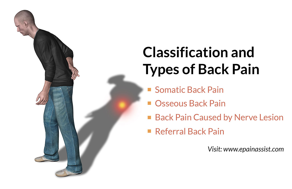
Classification and Types of Back Pain or Backache

Somatic Back Pain or Backache

  • Superficial: Localized in skin and subcutaneous tissue e.g. skin abscess, laceration, incision, contusion, hematoma and hair follicle infection.[1]
  • Deep: Localized in muscles and tendon e.g. muscle tear, muscle injuries, severe spasm, myofascial pain and fibromyalgia.[1]

Osseous Back Pain or Backache

  • Disc: Discitis, herniation and bulge.
  • Facet Joint: Arthritis, joint injury, joint hypermobility, joint instability and joint hypertrophy.
  • Vertebral Body: Metastatic tumor and fracture.
  • Foramina: Stenosis, fracture of pedicle and lamina, encroached by tumor mass.
  • Fracture: Body of vertebrae, transverse process and spinous process.[2]

Back Pain or Backache Caused by Nerve Lesion

  • Radiculopathy: Pinched nerve at foramina secondary to stenosis or tumor encroachment, bulged disc or herniated disc pinching the nerve.[3]
  • Spinal Stenosis: Spinal canal is narrowed secondary to facet joint hypertrophy, ligamentum flavum hypertrophy, disc bulge and herniation, subluxation of vertebrae due to tumor mass compromising the spinal canal.[4]

Referral Back Pain or Backache

Pain caused by diseases in distant organs may be felt (referred) in back (neck, mid back and lower back) as a backache. Many disorders within the abdomen, such as appendicitis, aortic aneurysms, kidney diseases, kidney infection, bladder infections, pelvic infections, and ovarian disorders among others, can cause pain referred to the back.[5]

References:

Also Read:

Back Pain And It’s Effects on Economy and Work Productivity

Back Pain And It’s Effect on Economy

In USA 65 million Americans have experienced back pain at least one time. Chronic back pain is observed in 16 million adults in USA at a given time. Healthcare cost incurred for investigation, lab study, treatment and disability payment is close to 12 billion dollars every year. Loss of income for not being able to work is close to 50 billion dollars per year. Back pain influences ability to work and also results in higher healthcare cost.(1)

Back Pain And It's Effect on Economy

Back pain in younger patient is caused by injury. Injury is caused by auto or work accident and domestic fall. In case of auto and work accident, cost of treatment is shared by auto insurance and workman’s compensation. Personal private insurance or Medicaid assumes cost of treatment of young individual following domestic fall if patient is uninsured. Back pain is also common among elderly population. Back pain in older patient is mostly caused by degenerative diseases. Medicare pays cost of treatment of elderly patients. Increased spending by Auto insurance and Workman’s Comp for treatment of back pain raises insurance premium. The increased cost is shifted to healthy individuals. Increased spending by Medicare and Medicaid for treatment of back pain is deducted from tax revenue.

Back Pain Effects on Work Productivity

Chronic back pain has major impact on work productivity.(2) Decline in work productivity has been observed secondary to sick leave, restricted work, and secondary gain due to injury. Up to 90% of people experience an episode of back pain without other health concerns and these symptoms will decrease or disappear on their own within one or two months. During this period patient may be referred to several physician and may undergo several investigations. Young and elderly suffer with back pain, though the cause of back pain is different. Back pain in younger population is caused by sports injury, automobile accident and work accident. Young population studies have indicated chances of early improvement and better early recovery by staying active and avoiding more than a few days of relative bed rest. According to the NHS (National Health Service)-UK, back pain is the largest cause of work-related absenteeism in the United Kingdom. Although back pain may be painful and uncomfortable, it is not usually serious and is not a life threatening disease. Cost of treatment for back pain includes cost of investigation, physician fees, hospital cost, lab studies and legal fees.

Treatment of back pain includes medications, chiropractic treatment, physical therapy, acupuncture, interventional pain therapy and surgery. Apprehensive patient may see multiple physicians and may demand several investigations to identify the cause of pain to his satisfaction. Demanding patient with low pain tolerance may drive cost of investigation and treatment higher. While patient with tolerance to pain and suffering may delay the treatment or may even avoid seeing multiple physicians, which will result in lower cost of treatment. Most of the cause of back pain is treated for symptomatic relief. The most unpleasant symptoms, which results from back disease along with pain are tingling, numbness and weakness in lower leg. Symptoms of tingling, numbness and weakness causes apprehension and worries. Patient demands the relief of associated symptoms hoping he or she may be paralyzed and bed ridden for rest of the life. Wide range of treatment choice available for back pain often results in trial of multiple treatments in short period of time thus increasing cost of treatment. Epidural injection is performed with and without hyaluronidase medications. Addition of hyaluronidase with cortisone had increased cost of treatment but valued benefits were not observed.1

Spending for investigation and treatment of back pain is much higher when compared to other common diseases. The reason for higher expenses is probably addition of cost of multiple treatments, which is provided to patient simultaneously and repeated several time. This retrospective study by Hart OR suggest higher resource use for management of back pain.2

The single therapeutic beneficial treatment for low back pain is yet unknown though scientist and medical specialist have tried several conservative and invasive treatment. Insurance providers have expressed their concern about increased cost of treatment of back pain, but avoided any research funding. American academy of pain medicine has expressed the concern of prevalence of chronic pain and annual cost in lost productivity. Patient and families have expressed the concern regarding loss of income and family hardship. The capital market drives on profit and loss. Profits are decreasing as salaries and bonuses have skyrocketed.

Medical equipment manufacturing companies, pharmaceutical industries and hospitals have increased the cost of their services and product. Insurance companies also want to increase profit by decreasing expenses. Expenses are shifted to patient by increasing co-pays and deductible. Insurance provider hopes patient will avoid or postpone the treatment. Insurance companies have created several layers for physician to get approval for lab or radiological studies and treatment.

Young male or female patients worry of back pain following auto, work or domestic accident. He or she is worried about loss of wages, job, how to pay for domestic expenses, co-pays, deductible and getting out of misery of back pain to go back to work. Physicians and hospitals are worried about how to get pre-authorization for investigations and treatment. Insurance companies are worried about increased cost of treatment and decreasing profit. Best outcome for young patient is to resume his work with no or less pain and for elderly patient is to continue with outdoor activities.

References:

  1. https://www.ncbi.nlm.nih.gov/pubmed/15062712
  2. https://www.ncbi.nlm.nih.gov/pubmed/15257978
  3. Epidural lysis of adhesions for failed back surgery and spinal stenosis: factors associated with treatment outcome.
    Hsu E1, Atanelov L, Plunkett AR, Chai N, Chen Y, Cohen SP. Anesth Analg. 2014 Jan;118(1):215-24.
  4. A study of National Health Service management of chronic osteoarthritis and low back pain.
    Hart OR1, Uden RM2, McMullan JE3, Ritchie MS4, Williams TD5, Smith BH6.
    Prim Health Care Res Dev. 2014 Mar 27:1-10.

Also Read:

Back Pain and It’s Effect on Elderly People, Young People, Drivers

Back Pain and It’s Effect on Elderly People

The population of elderly people in USA is increasing. Most of the working female and male are retiring at the age of 65 years. Most of the retirees had spent entire working life at work and hoped to travel or spend some time in out door sports following retirement. But majority of the older populations are now spending most of the time in taking care of back pain and other health related diseases. Majority of the back pain, about 98% or so, are nonspecific and self-limiting. Image studies have revealed that up to 85% of people with low back pain, despite a thorough medical examination, have not shown any specific cause of the pain. Symptoms may disappear within 2 to 4 weeks in 98% of the patients complaining of low back pain. Only 2% of patients continue having symptoms beyond 4 weeks and are supported by specific diagnosis. Back pain is the second most common symptom after upper respiratory tract infection to consult primary care physician in USA. It is also the second most common pain symptom after headache requiring frequent emergency room visits. Disc herniation has been observed in 33% of the adults older than 30 years, but only 2% suffer with long-term chronic pain. In USA, 90% of population have suffered with chronic low back pain and were unable to work for few days. After the initial episode of back pain, 50% of these individuals will have recurrent back pain at least once or more than once. Often radicular pain or sciatica pain is associated with low back pain. 80% of patients recover without surgery after suffering with just low back pain without radicular leg pain.

Back Pain and It’s Effect on Elderly People, Young People, Drivers

The study of 252 older patients suggests osteoarthritis of facet joint was the most common cause of back pain in older patients.5 Therapeutic management of back pain includes medications, physical therapy, interventional pain therapy and surgery. The guidelines and several options of treatment have resulted in increased cost of treatment. The cost of back pain management will increase as the aging population is also increasing. Low back pain is also common among obese patient6.

Back pain has restricted traveling and outdoor activities among retirees.

Back Pain And It’s Effect On Young People:

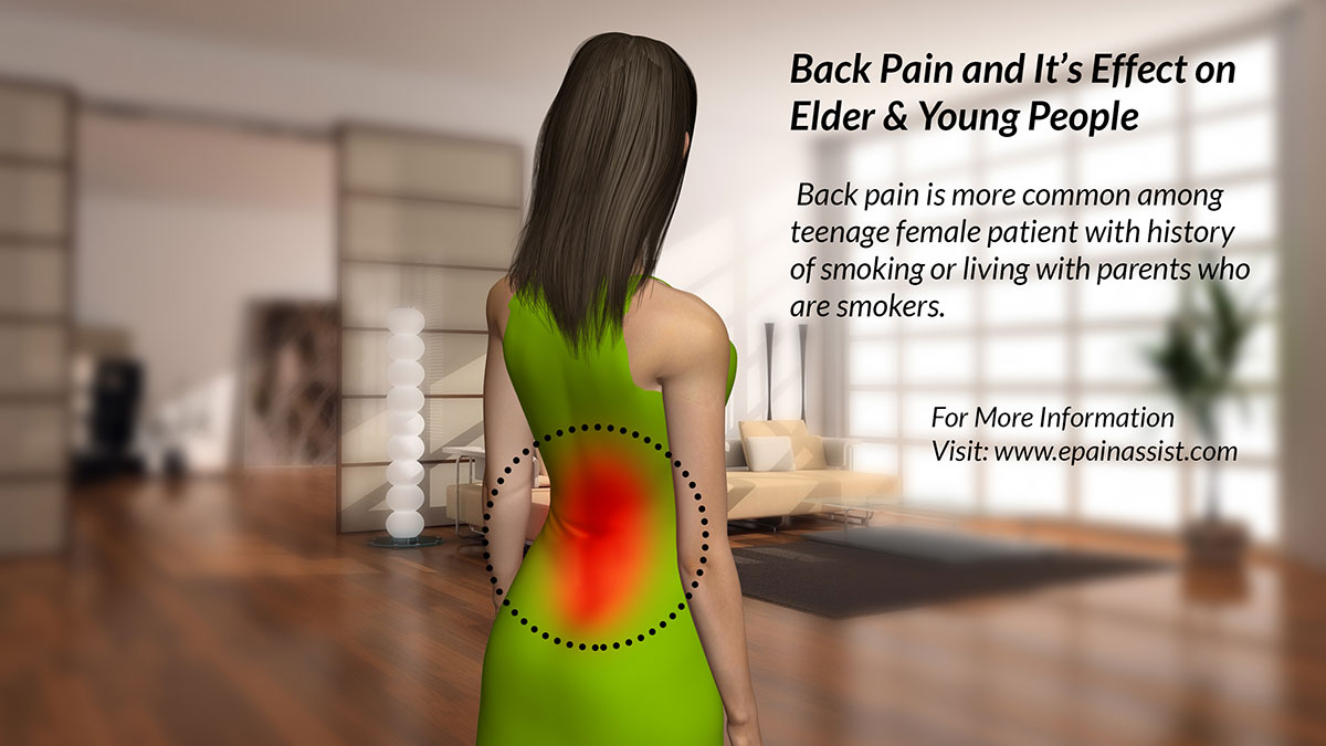
Recent published epidemiology study compared 4 levels of severe back pain. Study was published1 in 2014. Study suggests low back pain (LBP) causes more global disability than any other conditions.1 Chronic primary or secondary smoking causes back pain in younger population. Back pain is more common among teenage female patient with history of smoking or living with parents who are smokers.2 Back pain among younger workers was more related to work habit and working conditions. Proper posture and equipment had reduced the frequency of back pain among manual workers over the last two decades. Most of the back pain now is related to weight distribution and adopting posture while at work to prevent pain in extremities. Manual work causes extra load of weight transmitted through the back and posture during the work is adjusted by the worker to prevent pain in lower extremity. Study has suggested addition of extra sole in shoes prevent back pain.3

Back Pain And It's Effect On Drivers

Back Pain And It’s Effect On Drivers:

Low back pain is also common among professional drivers who may spend several hours in one position while driving either long distance or short distance with several breaks. Comparison between female and male driver suggests back pain is more common among female drivers than male drivers. Back pain among drivers is also related to the condition of the vehicle, which is driven regularly by same driver. Recent study of 8.4 million ethnically diverse US army drivers suggest risk of low back pain was higher among the driver than general population.4 Back pain is caused by abnormality or disease of muscles, vertebral column, epidural space and nerve pinch. Back pain in older population is often caused by degenerative disk or facet joint disease. Severe disk degeneration causes narrowing of disk height. Thinner disk causes foraminal stenosis resulting in pinch of spinal nerve or facet joint subluxation resulting in facet joint arthropathy. Pain caused by pinch nerve is often back pain with radicular pain and facet joint pain is mostly back pain.

Tips On Preventing Or Minimizing The Frequency Of Back Pain:

Past scientific studies suggest back pain can be prevented or frequency of back pain is minimized by avoiding smoking, maintaining proper posture during long time sitting, maintaining proper weight and performing regular exercise.

References:

  1. The global burden of low back pain: estimates from the Global Burden of Disease 2010 study.
    Hoy D1, March L, Brooks P, Blyth F, Woolf A, Bain C, Williams G, Smith E, Vos T, Barendregt J, Murray C, Burstein R, Buchbinder R.
    Ann Rheum Dis. 2014 Mar 24.
  2. Spine Day 2012: spinal pain in Swiss school children- epidemiology and risk factors.
    Wirth B1, Knecht C, Humphreys K.
    BMC Pediatr. 2013 Oct 5;13:159.
  3. The effect of cushioning insoles on back and lower extremity pain in an industrial setting.
    Jefferson JR
    Workplace Health Saf. 2013 Oct;61(10):451-7.
  4. Occupational driving as a risk factor for low back pain in active-duty military service members.
    Knox JB1, Orchowski JR2, Scher DL3, Owens BD4, Burks R5, Belmont PJ Jr3.
    Spine J. 2014 Apr 1;14(4):592-7.
  5. Presence and extent of severe facet joint osteoarthritis are associated with back pain in older adults.
    Suri P1, Hunter DJ, Rainville J, Guermazi A, Katz JN.
    Osteoarthritis Cartilage. 2013 Sep;21(9):1199-206.
  6. An epidemiologic analysis of low back pain in primary care: a hot humid country and global comparison.
    Bener A1, Dafeeah EE, Alnaqbi K, Falah O, Aljuhaisi T, Sadeeq A, Khan S, Schlogl J.
    J Prim Care Community Health. 2013 Jul 1;4(3):220-7.

Also Read:

Incapacitating Back Pain Due To Failed Back Syndrome

Incapacitating back pain is either acute or chronic in duration. Back pain may be localized over neck, mid back, lower back, and sacrum. A severe dreadful back pain along the vertebral column is also known as dorsalgia (back pain). Incapacitating chronic pain of neck, mid back, or lower back can originate from skin, subcutaneous tissue, tendons, muscles, cartilage, bones, and nerves.

Layers from skin to spinal cord are skin, subcutaneous tissue, supra-spinous ligament, inter-spinous ligament and posterior longitudinal ligament. Anterior to the spinal cord is anterior longitudinal ligament. There are several muscles attached to vertebrae. Muscles originate from vertebral body anteriorly, transverse process laterally, and spinous process posteriorly.

Disabling and dreadful chronic back pain also results following back surgery. Post surgical pain in the neck, mid back, or lower back is also known as failed back syndrome.(1)

Incapacitating Back Pain Due to Failed Back Syndrome

Failed back syndrome is caused by one of the following reason such as scar tissue pinching the nerves, weakness in facet joint, and traumatic injury of nerve. Healing of soft tissue after surgery can cause severe scarring of tendon, muscles or fatty adipose tissue. Scarring of adipose tissue, tendon, and muscles can cause pressure or pinching of surrounding nerve.

Back Pain Due To Failed Back Syndrome

Low back pain is more common than mid back pain or neck pain. Dreadful severe pain in any segment of vertebral column often causes short term or long-term disability.

Disability and Back Pain

After neck, mid back, or lower back injury there is often a question raised by individual suffering with chronic dreadful pain not responding to any treatment “Am I Disabled?” Answer is not simple. Disability is evaluated following optimization of treatment.(2) Treatment choice is medications, physical therapy, injection treatment, and surgery. Patient is considered disabled if treatments are not giving adequate pain relief or patient has permanent impairment.

Painless accomplishment of daily activities depends on correct alignment of vertebrae, muscles and ligaments during the mechanics of ambulation. Disparity in mechanics of weight bearing as well as joint movements such as flexion, extension, and rotation of vertebral column may result in chronic pain. Incapacitating back pain is the fifth most common reason to visit physician’s office. Even though back pain can affect people of any age, nonspecific benign backache causing incapacitating disability is significantly more common among working class adults aged between 35 and 55 years.

References:  

Also Read:

Tests to Diagnose Headache: Blood Tests, Imaging, EEG, Eye Examination, Spinal Tap

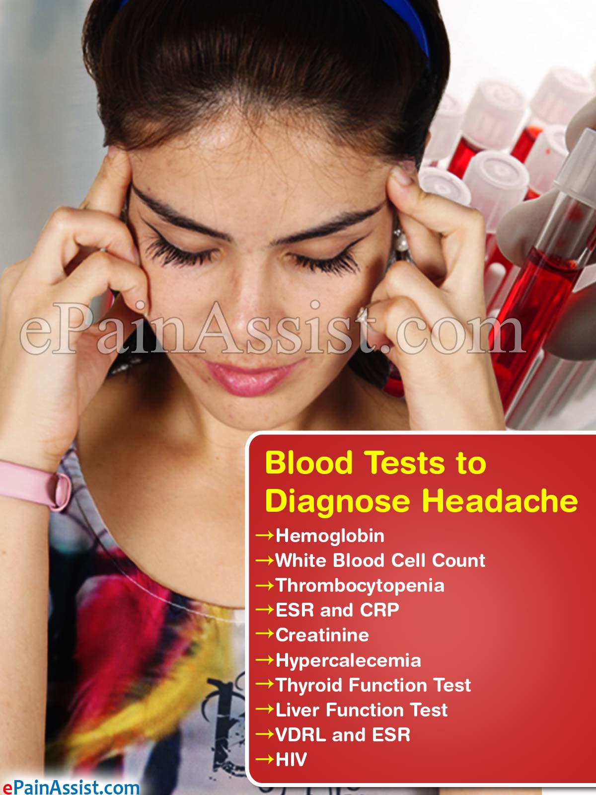
Blood Tests to Diagnose Headache

  • Hemoglobin – Low hemoglobin in anemia secondary bleeding or autoimmune disease.
  • White Blood Cell Count – Increased in infection e.g. meningitis, encephalitis.
  • Thrombocytopenia – Decrease platelet count in bleeding disorder causing headache.
  • ESR and CRP – Level increased in temporal arteritis or systemic disease.[1]
  • Creatinine – Kidney disease and kidney failure associated with headache.
  • Hypercalcemia – Increased calcium level in blood associated with headache.
  • Thyroid Function Test – Low thyroid hormone in hypothyroidism causing headache.[2]
  • Liver Function Test – Abnormal liver function test may be associated with primary or metastatic tumor of liver and brain.
  • VDRL and ESR – Increased in tuberculosis and veneral diseases associated with headache.
  • HIV – Patient with positive HIV may be suffering with headache.

Blood Tests to Diagnose Headache

Imaging Tests to Diagnose Headache

CT scan of brain – Useful in diagnosis of hemorrhage, brain tumor or brain metastasis.

Magnetic Resonance Imaging (MRI) of The Brain– Assist in evaluation and diagnosis of multiple diseases causing headache:

Contraindication for MRI

  • Pregnancy.
  • Kidney problem.
  • Sickle cell disease.
  • Devices – Arterial stent, pacemaker, mitral heart valve, spinal cord stimulator, intrathecal catheter and programmable pain pump, brain stimulator, limb prosthesis, IUD.
  • Remove metal object – jewelry, watches, Denture, hearing aids, hairpins, coins and credit cards.[4]

Sinus X-Ray to Diagnose Headache

Sinus infection such as frontal and maxillary sinusitis can cause sever headache. X-ray is used to diagnosed sinusitis.[5]

Electoencephalogram (EEG) to Diagnose Headache

Used in seizure disorder associated with headache.[6]

Eye Examination to Diagnose Headache

Eye examination with ophthalmoscope by eye specialist – rule out glaucoma and optic neuropathy.

Spinal Tap to Diagnose Headache

Spinal tap procedure is performed in operating room under sterile conditions. Cerebrospinal fluid is collected and studied for infection and autoimmune diseases.[7]

References:

Also Read:

Treatment for Headache: Medications, NSAIDs, Opioids, Oxygen Therapy, Surgery

Most individuals suffer from mild to severe headache at some point in their life. Headache is classified as tension headache, cluster headache, and migraine. Migraine is the most debilitating symptom and is observed in 4.4% of the patient visiting the office of General Practitioner.1 The tension headache is the most common type of headache and lasts for a few minutes to several hours.2 Most patients use over the counter medication to treat a tension headache. Most headaches are treated with NSAIDs and Tylenol. Occasionally lower dosage of opioids are prescribed for a headache that is resistant to most anti-headache medication. Among headaches, migraine resists most medication but frequently responds to triptans and ergotamine. The better therapeutic outcome is observed following treatment using triptan than ergots.3

Medication Treatment for Headache

Triptans

Sumatriptan is also known as a triptan. Triptan is prescribed for migraine and cluster headache. Triptans are prescribed as a nasal spray and intravenous therapy. Sumatriptan injection is very effective in the treatment of migraines and cluster headaches and works within 15 minutes. The nasal route of sumatriptan is effective in 30 minutes. In most of the cases, the effect of the nasal treatment lasts less than 45 minutes. Quality of pain relief is better with intravenous therapy than nasal spray treatment.3 Newer triptans with fewer side effects used are zolmitriptan (Zomig) in oral or nasal spray forms.

Treatment for Headache: Medications, NSAIDs, Opioids, Oxygen Therapy, Surgery

Side Effects of Sumatriptan Are As Follows:

  • Nausea.
  • Dizziness.
  • Muscle weakness.
  • Heaviness or pressure in the chest.
  • Tingling and numbness in the toes.
  • Rapid heart rate.

Complications of Sumatriptan:

Ergotamine

Dehydroergotamine (DHE) is prescribed for migraine and cluster headache. Most of the patients suffering from cluster headache respond within 5 minutes with no pain. Ergotamine can be prescribed as tablets, suppositories, and nasal spray.

Side Effects of Ergotamine:

  • Tachycardia.
  • Nausea and vomiting.
  • Diarrhea.
  • Dizziness.
  • Muscle weakness.
  • Heaviness or pressure in the chest.
  • Tingling and numbness in the toes.

Complications of Ergotamine:

  • Stroke secondary to vasoconstriction of cerebral arterial blood vessels.
  • Heart attack and angina secondary to vasoconstriction of the coronary artery.
  • Hypertension caused by vasoconstriction of the peripheral arterial system.

Serotonin syndrome is a life-threatening condition. Sumatriptan increases the overall concentration of serotonin in the brain. Symptoms observed are tachycardia, nausea, vomiting, diarrhea, hallucination, anxiety, tremors, and restlessness.

Lidocaine (Local Anesthetics)

Lidocaine– is a local anesthetic and used in resistant cases to treat a cluster headache. Lidocaine nasal drops or nasal spray is recommended for optimum effects.4 A group of patients with a history of cluster headache may not respond to lidocaine treatment. Reports suggest that it is helpful for most patients within about 40 minutes.

Side Effects of Lidocaine:

  • Unpleasant taste.
  • Numbness of area treated.

Capsicum for Headache

Capsicum originated from hot pepper, and it is used for tension and cluster headache. Capsicum is applied over occipital skin and also inside the nose. It is a safe medication and available as over the counter product in pharmacy.

Side Effects of Capsicum:

Serious Side Effects of Capsicum:

  • Liver and kidney damage and
  • Bleeding disorder.
  • It is recommended to discontinue medications for two weeks to prevent post-op bleeding if a patient is scheduled for surgery

Calcium Channel Blockers for Headache

Calcium channel blockers influence the calcium channel of the smooth muscle of blood vessels. A calcium channel blocker is effective in treating hypertension and coronary vascular disease. Migraine or sinus headache and cluster headache is secondary to changes in the diameter of cerebral blood vessels. Calcium channel blocker causes changes in the width of a cerebral blood vessel resulting in a reduction of headache symptoms. Verapamil is a preferred calcium channel blocker used for preventive treatment of cluster and migraine headache.5, 6 Medication should be taken for 2 to 3 weeks to build maximum effects.

Side Effects of Calcium Channel Blockers:

  • Constipation.
  • Severe Hypertension – If discontinued abruptly.
  • Bradycardia and Hypertension – If overdose.
  • Heart Failure – History of congestive heart failure.

Lithium for Headache

Lithium (Eskalith, Lithane, Lithobid, Lethonate, Lithotabs), commonly used for bipolar disorder, may also help prevent cluster headaches. The patient usually receives benefit within two weeks of starting to take the drug, often within the first week. Lithium may be used alone or with other medications. Lithium can have many side effects, including trembling of hands, nausea, and increased thirst. Weight gain is a common side effect with long-term use.

Side Effects of Lithium:

  • Thinning of the hair and hair loss.
  • Mild tremor.
  • Loss of appetite.
  • Tiredness.
  • Nausea.
  • Severe allergic reactions.
  • Skin – Rash, hives, itching.
  • Respiratory system – Bronchospasm, dyspnea and tightness in the chest.
  • Cardiovascular system – Slow or irregular heartbeat.
  • Central nervous system – Confusion, restlessness, tremor, dizziness, faintness, bladder and bowel incontinence, numbness of the skin, ringing in the ear, restlessness, seizures and severe or persistent headache.
  • Musculoskeletal system – Tremors, involuntary muscle twitching, decrease muscle tone, and coordination.
  • Gastrointestinal system – Edema of oral cavity and tongue, nausea, and diarrhea.

Anti-Seizure Medication Treatment for Headache

Anti-seizure medications are used for the prevention and treatment of headache.7 Anti-seizure medications thought to reduce the occurrence of migraines by calming inflamed blood vessels in the brain, which may lead to migraine pain or sinus headache. However, none of these medications is a cure for migraines. Antiepileptics are a diverse group of medications used as antiseizure. Some of the medication within this large group is being used to treat a headache. In research, there were areas of hyper activities in the brain that were recognized in epilepsy and headache patients. Antiepileptic medications reduced the area of irritability by its molecular action at the voltage-gated sodium channel. The most common used antiepileptic in headache treatment is Topamax, Depakote, and Neurontin.

Topamax – The oral tablet is taken every day as prescribed, even if headache symptoms are absent.

Side Effects of Topamax:

  • Tachycardia.
  • Dyspnea – breathlessness or shortness of breath.
  • Chest pain.
  • Urine – bloody or cloudy.
  • Cognitive symptoms – thoughts of suicide or aggressive behavior.
  • Depression.
  • Depakote – Depakote (valproic acid) is available as capsule, tablet and syrup.

Side Effects of Depakote:

Side Effects of Neurontin:

Nausea.

Serious Allergic Reaction:

  • Skin – Rash, hives, itching.
  • Respiratory System – Bronchospasm, dyspnea and tightness in the chest.
  • Central Nervous System – Seizures and Tremors.
  • Musculoskeletal System – Tremors.

Corticosteroids for Headache

Corticosteroid drugs (also called steroids) are used in allergic reaction and autoimmune diseases anti-inflammatory medications. Corticosteroids are membrane stabilizing agents, prescribed for the headache to prevent irritability of cortical and subcortical centers. It enhances the effect of verapamil in the treatment of headaches and often prescribed with verapamil. Prednisone is being tried as short term breakthrough pain meds for cluster headache. It is usually prescribed as a tablet form prednisone and injection form decadron.

Side Effects of Corticosteroids:

  • Symptoms like diabetes increase blood sugar levels.
  • General Symptoms – Dry mouth, fatigue, weakness, blurred vision, facial swelling, infection, avascular necrosis and glaucoma.
  • Central Nervous System – Anxious, depression, confusion, insomnia, mania, psychosis.
  • Gastrointestinal System – Stomach bloating feeling, increase appetite, weight gain, abdominal pain, diarrhea and peptic ulcer.
  • Urination – Frequent urination.

Nonsteroidal Anti-Inflammatory Drugs (NSAIDs) for Headache

  • NSAIDs are anti-inflammatory medication abbreviated as NSAIDs. NSAIDs are used for the treatment of headache in combination with other medications. Mild headaches are often treated by NSAIDs alone.
  • Analgesics – NSAIDs are not effective in severe pain and also pain caused by non-inflammatory etiology.
  • Most Prescribed NSAIDs – Aspirin, Ibuprofen (Motrin, Advil) and Naproxen (Aleve).
  • Side Effects – Complications caused by NSAIDs can be devastating if continued for a prolonged period.
  • Dose-dependent Side Effects – Dyspepsia, ulcer, perforation, upper gastrointestinal bleeding, and severe blood loss. A combination of aspirin and COX 2 inhibitor (Celebrex) may block the cardioprotective effects of aspirin.
  • Myocardial Infarction and Stroke – NSAIDs with COX2 inhibitor may increase risk. A combination of NSAIDs and COX 2 are not recommended in-patient with a history of myocardial infarction and heart attack.

NSAIDs Combinations With Other Medications Used In Headache

Most acute attacks of tension-type headaches get better without any treatment. Over-the-counter pain relievers such as acetaminophen or non-steroidal anti-inflammatory drugs (NSAIDs) can treat mild symptoms. Aspirin or ibuprofen (generic, Advil, other brands) are usually the first choices, followed by naproxen (generic, Aleve). Some patients may also find helpful medications that combine a pain reliever with caffeine.

  • Ibuprofen. (Advil, Motrin)
  • Naproxen. (Aleve, Naprelan, Naprosyn, Anaprox)
  • Acetaminophen/butalbital. (Bupap, Cephradine, Phrenilin)
  • Acetaminophen/butalbital/caffeine. (Esgic, Fioricet)
  • Aspirin/butalbital/caffeine. (Fiorinal)
  • Acetaminophen/butalbital/caffeine/codeine. (Fioricet with Codeine)
  • Acetaminophen/Dichloralphenazone/Sometheptene Mucate. (Midrin)
  • Acetaminophen/Phenyltoloxamine. (Percogesic)
  • Acetaminophen/Isometheptene mucate. (Prodrin)

Anti-Anxiety Medications for Headache

Sedatives are prescribed for selective cases of headache. The most common indication to use sedatives or anti-anxiety medications are Tension headache and prevent a recurrent episode. Buspirone (Buspar) and Alprazolam are prescribed as anti-anxiety medications.

Side Effects of Anti-Anxiety Medications:

  • Drowsiness.
  • Irritability.
  • Fatigue.
  • Low blood pressure.
  • Amnesia.

Opioid Medications for Headache

Opioids are used for treating tension headaches, which are not responding to NSAIDs and other medications. Acute and brief attack of headache most of the time gets better without any treatment. Patients may develop resistance and tolerance to opioid medication. Patients are advised to consider relaxation treatment, cognitive behavioral therapy, and lifestyle changes if failed to get pain relief with opioids. Daily attacks of headaches are treated with preventive medication such as NSAIDs, and NSAIDs combined with opiates or opioids. All other options of medications should be tried before introducing opioid prescriptions. Patients dependent on opioids for pain relief consume frequent pills for rapid pain relief and at the time may behave like a drug addict. Opioid dependence some time needs a higher dosage and frequent need for pain medications. These symptoms are because of opioid dependence to achieve pain relief and classified as Pseudoaddiction. Opioids do cause addiction in a few patients, and treatment of headache with opioids become difficult.

Side Effects of Opioids:

Common Side Effects of Opioids:

  • Nausea.
  • Vomiting.
  • Constipation.
  • Dry mouth.
  • Itching.
  • Miosis.

Less Common Side Effects of Opioids:

  • Skin – Itching, urticaria, hypothermia, and histamine-induced flushing.
  • Eyes – Miosis.
  • Gastrointestinal – Nausea, vomiting, dry mouth, constipation, and biliary spasm (colicky pain).
  • CNS – Depression, confusion, hallucination, delirium and dizziness.
  • Respiratory System.
  • Depression
  • CVS – Bradycardia and occasional may be tachycardia.
  • Orthostatic hypotension.
  • Kidney, Ureter, and Bladder (KUB) – Urinary retention and ureteric spasm.
  • Musculoskeletal – Muscle spasm.

Botox for Headache

Botulinum toxin A (Botox) injections are typically used to smooth wrinkles in the practice of plastic surgery. Botox is used for the treatment of headaches, including the prevention of cluster headaches.8 Research is still preliminary, and there is no sufficient evidence to support its efficacy. Botox is the treatment of choice for headache localized over frontal, occipital and cervical (neck) region. Headache over parietal (top of head), temporal area (side of head above the ear), between eyes and behind the ear does not respond to botox injection.

Frequent Common Side Effects:

  • General Symptoms – Irritated eyes, facial pain, flu-like symptoms, inability to focus the eyes, neck pain, redness, swelling, tenderness at the injection site; sensitivity to light, sweating, and tiredness.
  • CNS – Anxiety, arm or leg pain, back pain, dizziness, drowsiness, and headache.
  • Gastrointestinal System – Nausea, constipation, dry mouth, dry, and indigestion.
  • Respiratory System – Increased cough, mild sore throat, and runny nose.
  • Musculoskeletal – Stiff or weak muscles at or near the injection site.

Severe Allergic Reactions Need Immediate Medical Attention:

  • Skin – Swelling of the mouth, face, lips, or tongue; fever and chills.
  • Respiratory system – Unusual hoarseness, wheezing, chest pain, difficulty swallowing or breathing, persistent sore throat and shortness of breath.
  • Kidney Ureter Bladder – Blood in the urine; burning, numbness, or tingling; burning with urination; difficult, frequent, or painful urination, loss of bladder control, eyes-double or blurred vision, or other vision changes; drooping of the upper eyelid and eyelid swelling.
  • Central Nervous System – Fainting; seizures, headache, dizziness speech changes or problems and worsening migraines.
  • Cardiovascular System – Irregular heartbeat.
  • Musculoskeletal System – Loss of strength, paralysis, and severe or persistent muscle weakness.

Melatonins-(N-acetyl-5-methoxytryptamine) for Headache

Melatonin is found in animals, plants, and microbes. Melatonin is sold over the counter in the USA. In an animal research, melatonin was investigated against carcinogens. Melatonin prevents the initiation of the impairment of DNA by carcinogens. Impaired DNA otherwise would have initiated cancer growth.

Melatonin is being prescribed for migraines, as well as tension headache.9 Melatonin is widely used as an antioxidant in diseases such as immunodeficiency syndrome, autism, aging, Alzheimer disease, delirium mood disorder, alternative medicine, tinnitus, cancer and gallstone.

Oxygen Therapy for Headache

Oxygen therapy – Oxygen percentage in the atmosphere is 21%. Oxygen therapy can provide from 30% to 100% oxygen. Nasal oxygen therapy using nasal cannula offers 30 to 40% oxygen. A tight-fitting face mask or endotracheal tube can deliver up to 100% oxygen. Oxygen therapy has been used in congestive heart failure, chronic obstructive lung disease (COPD), and allergic reaction causing shortness of breath, convulsion, and hemorrhagic shock. Oxygen therapy up to 100% is used in cluster and tension headache on and often. The patient is advised to inhale oxygen for 15 minutes several times a day.

Surgery for Headache

  • Occipital Neuralgia- Pain over the back of the head (occipital area of a skull) is caused by irritation of the lesser and greater occipital nerve. Such pain is treated with radiofrequency ablation of bother lesser and greater occipital nerve.10
  • Deep Brain Stimulation: Chronic cluster and migraine headaches in a few patients are resistant to all treatments. These patients are imprisoned in the house suffering from an intense headache and unable to perform any regular activities. Severe pain may bring multiple thoughts of suicide. These patients are given a trial of deep brain stimulation as a last option of treatment. The published studies suggest that implants of the hypothalamic stimulator have a better outcome in treating a cluster headache.11
  • Gasserian (Trigeminal) Ganglion Radiofrequency Rhizotomy: Headache associated with trigeminal nerve irritation or pinch is treated with Gasserian ganglion neurotomy. Gasserian or trigeminal ganglion ablation is performed by using radiofrequency heat12 or glycerol. The trigeminal ganglion is identified during the procedure by sensory nerve stimulation. The trigeminal ganglion is treated with heat that is produced by radiofrequency waves. Pain carrying fibers are also often destroyed in few cases using glycerol as a chemical rhizotomy.
  • Microvascular Decompression of Trigeminal Nerve: Trigeminal nerve pain is observed in patients suffering from postherpetic neuralgia and traumatic injury of the trigeminal nerve. Chronic headache pain secondary to trigeminal neuralgia is treated with microvascular decompression of the trigeminal nerve.13 Blood vessels surrounding the trigeminal nerve are selectively ligated or cauterized. Such treatment eliminates the pressure over the trigeminal ganglion.

Craniotomy for Headache

A craniotomy is a surgical procedure performed to the exposed brain and subcortical part of the brain. It is a highly skilled procedure, which involves the removal of part of the skull bone and reattached at the end of surgery. Surgery is performed under the guidance of a CT scan. MRI images are studied before surgery as well as during operation.

Indication for Craniotomy Procedure:

  • Tumor and Metastasis – Excision.
  • Aneurysm – Clipping and reanastomosis.
  • Blood Clot – Evacuation and stoppage of bleeding.
  • Brain Abscess – Excision and drainage.
  • Skull Fracture – Repair.
  • Ischemic Brain Tissue – Excision.

Risk Involved:

  • Infection.
  • Bleeding.
  • Seizures.
  • Dural tear – CSF leak.
  • Neurological Deficit – sensory and motor nerve deficiency, paralysis.
  • The blood clot, thrombosis, and embolism.
  • Cognitive Deficiency – memory, emotional expression.

Cognitive Behavioral Treatments for Headache

Cognitive Behavior Therapy (CBT) is useful to assist change or modify behavior problems in patients suffering from chronic headaches or sinus headaches.

Following modification of behavior helps to reduce or eliminate headache:

Behavioral Treatment Is Classified As Follows:

  • Cognitively Oriented.
  • Behaviorally Oriented.
  • Imaginary Exposure Therapy Combines Both Approaches.
  • Principle of Behavioral Treatment:
  • Points out over generalizing of symptoms.
  • Magnifying negative feelings.
  • Minimizing positive thoughts.
  • Guiding to more realistic, effective concepts.
  • Assess, reconceptualized, and acquisition of positive skills.

The Goal of Behavioral Therapy:

Modify Undesired Behavior – The treatment is targetted to eliminate and reinforced the changed behavior. Such therapy is performed as individuals or as a group.

Therapy as a Treatment – CBT has also been used to treat a primary and secondary headache that is causing severe pain and not responding to any alternative treatment. CBT is an active treatment inpatient suffering from headaches and insomnia.

Combination of Behavioral Therapy with Cognitive Therapy- Several centers have tried simultaneous behavior and cognitive therapy that has provided better results.

Maladaptive Thinking- Maladaptive thinking causes stress to induce a headache. Behavioral therapy helps to modify or eliminate maladaptive thinking.

Systemic Desensitization –

Systematic desensitization involves gradually exposing a patient to anxiety-provoking stimuli until the anxiety response was extinguished, or eliminated.

Positive Reinforcement

Positive reinforcement improves behavior when compare with conditioning therapy. The therapy recommends punishment for repetitive harmful behavior and rewards for improvement in behavior.

Behavioral therapies can be a helpful accompaniment to drug treatment. These approaches can help with pain management and enable patients to feel more in control of their condition.

References:  

  1. The diagnosis and treatment of chronic migraine Mark W. Weatherall, Ther Adv Chronic Dis. 2015 May; 6(3): 115–123. 
  2. Tension type headache Debashish Chowdhury, Ann Indian Acad Neurol. 2012 Aug; 15(Suppl 1): S83–S88.
  3. Triptans in the Acute Treatment of Migraine: A Systematic Review and Network Meta-Analysis. Cameron C1,2, Kelly S2, Hsieh SC2, Murphy M2, Chen L2, Kotb A2, Peterson J2, Coyle D1, Skidmore B2, Gomes T3, Clifford T1,4, Wells G1,2., Headache. 2015 Jul-Aug;55 Suppl 4:221-35.
  4. Intranasal lidocaine for cluster headache. Robbins L1., Headache. 1995 Feb;35(2):83-4.
  5.  Verapamil for cluster headache. Clinical pharmacology and possible mode of action. Tfelt-Hansen P1, Tfelt-Hansen J., Headache. 2009 Jan;49(1):117-25. doi: 10.1111/j.1526-4610.2008.01298.x.
  6.  Clin Neuropharmacol.1986;9(4):311-28. Calcium channel antagonists and the treatment of migraine. Greenberg DA.
  7. Antiepileptic drugs: how they work in headache. Cutrer FM1., Headache. 2001 Nov-Dec;41 Suppl 1:S3-10.
  8. Botulinum toxin in the management of chronic migraine: clinical evidence and experience Claus M Escher, Lejla Paracka, Dirk Dressler, and Katja Kollewe, Ther Adv Neurol Disord. 2017 Feb; 10(2): 127–135.
  9. The therapeutic potential of melatonin in migraines and other headache types. Gagnier JJ1., Altern Med Rev. 2001 Aug;6(4):383-9.
  10. Treatment of Occipital Neuralgia by Thermal Radiofrequency Ablation Lance M. Hoffman, MD,1 Alaa Abd-Elsayed, MD, MPH,2 Tim J. Burroughs, MD,1 and Harsh Sachdeva, MD1 , Ochsner J. 2018 Fall; 18(3): 209–214.
  11. Deep brain stimulation in headache. Leone M1., Lancet Neurol. 2006 Oct;5(10):873-7.
  12. Long-term results of radiofrequency rhizotomy in the treatment of cluster headache. Taha JM1, Tew JM Jr., Headache. 1995 Apr;35(4):193-6.
  13. Microvascular Decompression for Trigeminal Neuralgia: A regional unit’s experience Cathal Hannan, Alireza Shoakazemi, and Gavin Quigley, Ulster Med J. 2018 Jan; 87(1): 30–33.

Also Read:

Preventive Treatment for Headache: Cluster, Migraine, Tension Headaches

Preventing Attacks of Cluster Headache

Attacks are usually short, lasting from 15 – 180 minutes, and the excruciating pain may have subsided by the time a patient reaches a doctor’s office or emergency room. Because it can be difficult to treat attacks when they occur, treatment efforts focus on the prevention of attacks during cluster cycles.[1] Although certain drugs are standard, preventive therapy needs to be individually tailored for each patient. The doctor may prescribe a combination of drugs.

Verapamil (Calan), a calcium-channel blocker drug, is the mainstay preventive treatment for cluster headaches. However, it can take 2 to 3 weeks for this drug to take effect. During this period, corticosteroids (typically prednisone) may be used as an initial transitional therapy. For long-term treatment of chronic cluster headaches, lithium may be used as an alternative to verapamil.[2]

Although they are not approved for cluster headache, anti-seizure drugs such as valproate (Depakote), topiramate (Topamax), and gabapentin (Neurontin), are sometimes used for preventive treatment.

Headache Preventive Treatment

Preventing Attacks of Migraine Headache

Several medications are used for prevention of migraine.

  • Beta Blockers – Inderol and Toprol, which relax blood vessels.
  • Calcium channel blockers – Cardizem and Procardia, vasodilator and relaxes blood vessels causing migraine headache.
  • Tricyclic antidepressants – Amitriptyline.
  • Anticonvulsants – Depakote and Topamax.[3]

Preventing Attacks of Tension Headache

Avoid anxiety, fatigue, and hypoglycemia which triggers tension headache. Preventive medications used in tension headaches are:

  • Tricyclic antidepressants – Amitriptyline.
  • Anticonvulsants – Depakote and Topamax.
  • NSAIDs – Advil and Aleve.[4]

References:

Also Read:

Diagnosis of Headache: Migraine, Tension, Cluster, Stroke and TIA

Diagnosis of Migraine Headache

Migraine headache is severe intense throbbing pain with pulsating character mostly localized on one side of head. Patient may suffer with nausea and vomiting during the episode of headache. Patients are sensitive to light and sound. Pain may last for hours or days. Migraine headache may precede or accompany aura. Auras are sensory symptoms such as light flashing, blind spot or feeling of tingling and numbness.[1] Aura may precede migraine and considered as warning symptoms. Physical activities increase intensity of headache.

Diagnosis of Headache: Migraine, Tension, Cluster, Stroke and TIA

Diagnosis of Tension Headache

Tension headache may be acute in nature or rarely become chronic with daily pain lasting more than 3 to 6 months. Tension headache may be once or few times a week or continuous for a several days. If tension headache last for 15 days or more in one month it is considered chronic tension headache.[2] Headache is mild to moderate in intensity compared to migraine or cluster headache. Tension headache is not preceded or accompanied by aura like symptoms. Physical activities do not increase intensity of headache. Headache is either unilateral or bilateral.

Diagnosis of Tension Headache

Diagnosis of Cluster Headaches

Cluster headaches are one of the most painful cyclical headaches. Pain is often around the eyes and patient may wake up often in the middle of the night. Headache is unilateral like migraine headache. Cluster period of attack may last from weeks to months. In between attacks patient may pass through remission period of months to year. It is one of the rare types of headache.[3]

Diagnosis of Cluster Headaches

Diagnosis of Stroke and Transient Ischemic Attack (TIA)

Migraine patients with a history of frequent auras or cluster headaches may exhibit history of aura in 14% cases.

TIA is transient lack of blood supply to brain for very short period of time and may not induced brain damage or serious ischemia. But, if sustained for longer time may cause brain injury or stroke because of lack of blood supply (ischemia) to brain tissue. Persistent and frequent TIA may result in brain injury or stroke. TIA symptoms are brief and similar to those of stroke.[4] TIA is often a warning sign indicating a person is at risk for severe stroke. Headaches with auras may also increase the risk for eye retinal damage (retinopathy). Aura-related headaches may affect the small blood vessels in the brain and the eyes, thereby increasing the risks for stroke and retinopathy.[5] Cluster headaches are rare, affecting less than 1% of the population.

Pathophysiology causing these events and how they relate to cluster headaches are still unclear.

References:

Also Read:

Signs and Symptoms of Headache & How Long Do Headache Symptoms Last? | Identifying the Type of Headache

Pain in the forehead region and the scalp are the common symptoms of headache. Most cases of headache and its accompanying symptoms do not require immediate medical attention but if the frequency of headache is more accompanied by severe pain in the head, or after a trauma to the head and neck region or is accompanied with SOB, stiff neck, fever then it is better get yourself evaluated by an headache specialist at the earliest.

Symptoms of Migraine Headaches

Pain Intensity Evaluation of Headache Based on the Symptoms:

  • Symptoms of Mild Intensity Headache – Barely noticeable.
  • Symptoms of Moderate Intensity Headache- Does not interfere with work or activities.
  • Symptoms of Severe Headache- Distracts from work or activities.
  • Symptoms of Very Severe Headache- Difficult to maintain normal activities.
  • Symptoms of Incapacitating Headache- Unable to concentrate, needs to go to emergency room or see physician immediately.[1]

Symptoms of Migraine Headaches

How Long Do Headache Symptoms Last?

Frequency of Headache Symptoms:

  1. Symptoms of low frequency headaches last less than 15 days in a month.
  2. Symptoms of high frequency headaches last more than 15 days.[2]

Duration of Headache Symptoms:

  1. Short duration headache symptoms last less than 4 hours.
  2. Symptoms of long duration headache last more than 4 hours.

Diagnosis of Type of Headache Based on Frequency and Duration of Its Symptoms

Low Frequency and Long-Duration Headache Could be a Sign of:

  1. Migraine
  2. Episodic Tension Type Headache.

High Frequency and Long-Duration Headaches Could Be a Sign of:

Low Frequency and Short Duration Headache Could be a Sign of :

  1. Episodic Cluster Headache (not triggered by cough or exercises)
  2. Valsalva or Headache Triggered By Cough, Exercises or Valsalva

High Frequency and Short Duration Headache Could be a Sign of:

  1. Chronic Cluster Headache.
  2. Chronic Paroxysmal Hemicrania.
  3. Hypnic Headache.
  4. SUNCT Syndrome.
  5. Valsalva or Headache Triggered by Cough, Exercises or Valsalva.[3]

Identifying the Type of Headache Based on Age and Gender

  • Headache in young adults of age around 40 is more often a sign of cluster headache.
  • Men are more likely to suffer from more common cluster headaches, it is 2 to 3 times more often than women.
  • Headache in women aged 60 or more, is more likely a sign of Migraine headaches.[4] This type of headache is influenced by fluctuations in estrogen and other female hormones in females.

Identifying the Type of Headache Based on Lifestyle and Personal History

  • Symptoms of Headache in Nicotine Dependent (smoker) can be a sign of Cluster Headache.
  • Symptoms of Headache in Alcohol Abuser can also be a sign of Cluster Headache.
  • Professional Stress – Sign of Tension Headache and Cluster Headache.
  • Symptoms of headache after drinking red wine can be indicative of Cluster headache.
  • Discontinuation of smoking or drinking alcohol does not eliminate attacks of headache once it is established.

Identifying the Type of Headache Based on Medical History & Following Signs and Symptoms

  • Headache accompanied with Swollen Eyes and Lacrimation can be a sign of Cluster headache.
  • Headache accompanied with running nose and lacrimation – Cluster headache.
  • Headaches accompanied with Aura can be a Migraine.
  • Headache after Physical Activities can also be indicative of Migraine.
  • Bilateral Headache – Tension headache.

Type of Headache Based on Family History and Genetic Factors

Cluster and migraine headaches tend to run in families, suggesting a genetic component may be involved in some cases. About half of people with migraine headache have a personal or family history of migraine. Studies have reported that about 15% of patients have both kinds of headache.[5]

Type of Headache Based on Head Injury

Type of Headache Based on Sleeping Disorders

Identifying the Type of Headache Based Signs of Sleeping Disorders

Cluster headaches have been observed during specific sleep stages. Sleep disorders such as sleep apnea and narcolepsy tend to be followed by cluster headache or migraine headache. Patients who are unable to sleep (insomnia), commonly tend to suffer with cluster or migraine headache.[6] Sleep hypoxia secondary to sleep apnea may trigger physiological events causing cluster headache during sleep. Sleep hypoxia triggered cluster headache may continue its symptom behavior during daytime mostly during late afternoon. Most of the patient with history of sleep apnea causing cluster headache have responded well to the treatments like CPAP (continuous positive airway pressure ventilation). Sleep study is extremely important investigation in patient with history of cluster headache that is not responding to medication treatment.

References:

Also Read:
Pathophysiology of Headache: Primary Headaches, Secondary Headaches

Primary Headaches:

Primary headaches are migraine, cluster and tension headache. Primary headaches are benign, chronic and not life threatening. Headache is considered primary when a disease or other medical condition does not cause the headache. Most primary headaches fall into three main types: tension-type, migraine, and cluster headaches. Tension headache is the most common primary headache and accounts for 90% of all headaches. After tension headache, the second most frequently occurring primary headaches are Migraine. Migraine is referred to as a neurovascular headache because it is most likely caused by an interaction between blood vessel and nerve abnormalities. Cluster headache is a less common type of primary headache. Cluster headache sometimes referred to as a neurovascular headache, evidence now suggests that its cause may lie in the hypothalamus, a region deep in the brain that regulates, among other functions, the biologic rhythms of the body.[1]

Pathophysiology of Headache: Primary Headaches, Secondary Headaches

Secondary Headaches:

Secondary headaches are sometimes life threatening, may be secondary to metastasis or malignant cancer and cerebral bleeding. Early diagnosis of secondary headache is essential to avoid life-threatening complications. Secondary headache may be resulting from life threatening disease such as brain tumor, brain hemorrhage or head injury. Secondary headaches are classified based on etiology, not on symptom profile. Recent origin headache in a patient with history of disease known to cause intracerebral complications must be further investigated. Patients with pre-existing primary headaches sometimes experience exacerbations, which may follow further expensive investigations to rule out life threatening secondary headaches. In this circumstance, the known cause of headache may have worsened the pre-existing primary headache. Alternatively, it may have caused a new type of secondary headache. Secondary headaches are caused by other medical conditions, such as sinus infections, neck injuries, and strokes. About 2% of headaches are secondary to abnormalities or infections in the nasal or sinus passages, and they are commonly referred to as sinus headaches.

Traumatic injury to head and neck may cause headache which is classified as secondary headache. Head injury may cause subdural or epidural hematoma if ignored can lead to cerebral herniation and critical outcome of complications. Neck injury following work accident or auto accident causes occipital neuralgia and occipital headache. Intracranial arterial aneurysm or cerebral arterial swelling can cause severe throbbing headache. Infection such as meningitis or encephalitis can cause pain mostly in frontal and occipital area of head. In few cases of inflammatory disease of eye, ear, nose and sinuses pain simulating headache referred to temporal, frontal or occipital region. Shingles or post herpetic neuralgic pain of trigeminal nerve or facial nerve can also cause severe headache in frontal or temporal dermatome.[2]

References:

Also Read:

Classification and Types of Headache: Primary Headaches, Secondary Headaches

International Headache Society (IHS) first classified different types of headache in 1988. Headache classification were modified and improved in 2004 by International Headache Society. ICHD-2 classification of 2004 has simplified symptomatic descriptions and causation of headaches in to several subgroups, which were overlapping in the previous classifications.

Types Of Headache-Primary and Secondary

The ICHD- 2 Classification of Headache

  • ICHD has included almost all causes and symptomatic presentation of headache as symptoms.
  • Like its predecessor, the ICHD-2 separates headache into primary and secondary disorders.
  • ICHD-2 classification helps to achieve methodical approach to investigate and rule out symptoms with red flags causing secondary headache.[1]

Headache Is Classified In Two Main Groups as Follows

  1. Primary Headache.
  2. Secondary Headache.

A. Primary Headaches-

  • Primary headaches are benign, chronic and not life threatening.
  • Headache is considered primary when a disease or other medical condition does not cause the headache.
  • Most primary headaches fall into three main types: Tension-type, Migraine, and Cluster Headaches.
  • Tension headache is the most common primary headache and accounts for 90% of all headaches.
  • After tension headache, the second most frequently occurring primary headaches are Migraine.
  • Migraine is referred to as a neurovascular headache because it is most likely caused by an interaction between blood vessel and nerve abnormalities.
  • Cluster headache is a less common type of primary headache.
  • Cluster headache is sometimes referred to as a neurovascular headache, evidence now suggests that its cause may lie in the hypothalamus, a region deep in the brain that regulates, among other functions, the biologic rhythms of the body.[2,3]

Types of Primary Headache-

  1. Migraine Headache.
  2. Tension Headache
  3. Cluster Headache.

B. Secondary Headaches

  • Secondary headaches are sometimes life threatening, may be secondary to metastasis or malignant cancer and cerebral bleeding.
  • Early diagnosis of secondary headache is essential to avoid life-threatening complications.
  • Secondary headache may be resulting from life threatening disease such as brain tumor, brain hemorrhage or head injury.
  • Secondary headaches are classified based on etiology, not on symptom profile.
  • Recent origin headache in a patient with history of disease known to cause intracerebral complications must be further investigated.
  • Patients with pre-existing primary headaches sometimes experience exacerbations, which may follow further expensive investigations to rule out life threatening secondary headaches. In this circumstance, the known cause of headache may have worsened the pre-existing primary headache.
  • Alternatively, it may have caused a new type of secondary headache.
  • Secondary headaches are caused by other medical conditions such as sinus infections, neck injuries, and strokes.
  • About 2% of headaches are secondary to abnormalities or infections in the nasal or sinus passages, and they are commonly referred to as sinus headaches.[3]
  • Traumatic injury to head and neck may cause headache, which is classified as secondary headache.
  • Head injury may cause subdural or epidural hematoma and if ignored can lead to cerebral herniation and critical outcome of complications.
  • Neck injury following work accident or auto accident causes occipital neuralgia and occipital headache.
  • Intracranial arterial aneurysm or cerebral arterial swelling can cause severe throbbing headache.
  • Infection such as meningitis or encephalitis can cause pain mostly in frontal and occipital area of head.
  • In few cases of inflammatory disease of eye, ear, nose and sinus pain simulating headache is referred to temporal, frontal or occipital region.
  • Shingles or post herpetic neuralgic pain of trigeminal nerve or facial nerve can also cause severe headache in frontal or temporal dermatome.[4]

Types of Secondary Headache:

a. Traumatic Headache-

b. Vascular Headache-

c. Space Occupying Lesions-

  • Malignant tumor
  • Metastatic brain tumor
  • Brain abscess
  • Brain hemorrhage and blood clots[6]

d. Withdrawal Headache-

e. Infection-

f. Cerebral Ischemia

  • Embolism of cerebral blood vessels causes stroke and secondary headache.
  • Arterial spasm causes Transient Ischemic Attack or TIA.[7]

g. Psychiatric Disorders.

  • Depression may be associated with headache
  • Anxiety is often followed by headache.

h. Occipital Headache

  • Occipital headache is caused by vision problems.
  • Cataract or retinopathy may cause occipital headache.
  • Diplopia or vision problems results in severe headache.

References:

Also Read:

What is Headache or Cephalalgia?

What is Headache or Cephalalgia?

Headache or Cephalalgia is a symptom of pain observed in head, face and upper cervical dermatome. Headache or Cephalalgia is a complaint of discomfort originating from tissue covering brain such as meninges, skull, periosteum and structures of the skull base. In addition, pain also originates from blood vessels and muscles around brain, scalp, face, and neck. Cerebral cortex, cerebellum, midbrain, blood vessels and nerves occupy space within the skull.[1] Brain tissue is insensitive to pain since pain receptors are not detected in brain tissue.

Headache or Cephalalgia is broadly classified as primary and secondary headache.[2]

Migraine, cluster and tension headache are primary headache.

Space occupying lesion within skull causes secondary headache. Stretching of meninges by tumor, abscess and blood clot results in tension type secondary headache.

Headache

Secondary tension headache may incur because bony skull cannot expand with space occupying lesions. Increased tissue mass either tumor, abscess or blood clot within skull will cause increased pressure or compression of nerves and blood vessels. Occasional symptom of tension headache may be a warning sign of life threatening internal bleeding within compact bony skull.

Bleeding could be mild, moderate or severe and result in blood clot of variable size depending on blood volume. Initial symptoms of mild to moderate headache or Cephalalgia secondary to mild hemorrhage if ignored may result in continuation of internal bleeding and developing into massive blood clot. Massive blood clot will cause shift of cerebral vital centers towards the skull base and mid brain to the base of the skull.

Increased pressure and ischemia of vital brain centers will lead to life threatening symptoms such as slow breathing, slow heart rate, low blood pressure and respiratory apnea. Bleeding within brain tissue during initial phase may indicate mild to moderate headache or Cephalalgia.[3] Later with time, continuous slow bleeding may cause severe headache with substantial increase in size of clot. In a few cases, time is essential and important to save life.

References:

Also Read:

Differential Diagnosis for Abdominal Pain

What is the Differential Diagnosis for Abdominal Pain?

What is the Meaning of the Term Differential Diagnosis?

The procedure of separation between two or more illnesses which have comparable signs or indications is called as differential diagnosis.[1]

What is the Differential Diagnosis for Abdominal Pain?

Differential Diagnosis for Abdominal Pain

While diagnosing an individual with abdominal pain of acute nature, the doctor usually concentrates on regular conditions that bring about stomach pain or abdominal pain and additionally on more severe conditions. The point of pain or the location of the abdominal pain will usually help in diagnosis. For example, when diagnosing a patient with appendicitis, the location of the pain can give a good hint in the diagnosis of the condition.

Differential Diagnosis for Abdominal Pain or Stomach Ache

An ultimate diagnosis is not generally done at the initial visit of the patient; accordingly, it is basic to start the assessment by dismissing out severe conditions for example vascular illnesses like mesenteric ischemia and aortic dissection and surgical problems such as cholecystitis and appendicitis. The doctors will also examine the wall of the abdomen related conditions like herpes zoster, muscle strain in light of the fact that these are regularly misdiagnosed.[2]

Differential Diagnosis of Abdominal Pain Based on the Location

If the location of the pain is in the Right Upper Quadrant of the abdomen, the potential diagnoses are specified below:

  • If the location of the pain is detected in the biliary region of the upper right quadrant then the potential conditions could be cholelithiasis, cholangitis or cholecystitis.
  • If the pain location is detected in the colonic region of the upper right quadrant then the potential conditions could be diverticulitis or colitis.
  • If the location of the pain is detected in the Hepatic region of the upper right quadrant then the potential conditions could be hepatitis, a mass or abscess.[3]
  • If the location of the pain is detected in the pulmonary region of the upper right quadrant then the potential conditions could be embolus or pneumonia.
  • If the location of the pain is detected in the renal region of the upper right quadrant then the potential conditions could be pyelonephritis or nephrolithiasis.

In case the pain or the affected area is near the Epigastric region of the abdomen, then the conceivable findings are given below:

  • If the pain location is detected in the biliary location of the epigastric region of the abdomen then the potential conditions could be cholangitis, cholecystitis or cholelithiasis.
  • If the location of the pain is detected in the cardiac location of the epigastric region of the abdomen then the potential conditions could be pericarditis or myocardial localized necrosis.
  • If the pain location is detected in the gastric location of the epigastric region of the abdomen then the potential conditions could be peptic ulcer, gastritis or esophagitis.
  • If the pain location is detected in the pancreatic location of the epigastric region of the abdomen then the potential conditions could be a mass or pancreatitis.
  • If the pain location is detected in the vascular location of the epigastric region of the abdomen then the potential conditions could be mesenteric ischemia or aortic dissection.[4]

If the pain area is near the Left upper quadrant of the abdomen, the conceivable findings are given beneath:

  • If the location of the pain is detected in the cardiac region of the upper left quadrant then the potential conditions could be pericarditis, myocardial localized necrosis or angina.
  • If the pain location is detected in the gastric region of the upper left quadrant then the potential conditions could be peptic ulcer or esophagitis or gastritis.
  • If the location of the pain is detected in the pancreatic region of the upper left quadrant then the potential conditions could be pancreatitis or a mass.
  • If the location of the pain is detected in the renal region of the upper left quadrant then the potential conditions could be pyelonephritis or nephrolithiasis.
  • If the location of the pain is detected in the vascular region of the upper left quadrant then the potential conditions could be mesenteric ischemia or aortic dissection.[5]

In case patient feels that the pain is near the Periumbilical area of the abdomen, the conceivable determinations are given below:

If the pain location is detected in the colonic location of the periumbilical region of the abdomen then the potential conditions could be an early appendicitis.

If the pain location is detected in the gastric location of the periumbilical region of the abdomen then the potential conditions could be small bowel mass or blockage, gastritis, peptic ulcer or esophagitis.

If the pain location is detected in the vascular location of the periumbilical region of the abdomen then the potential conditions could be mesenteric ischemia or aortic dissection.

If patient complains about discomfort in the Right lower quadrant of the abdomen, the conceivable findings are given beneath:

  • If the location of the pain is detected in the colonic region of the lower right quadrant then the potential conditions could be colitis, appendicitis, diverticulitis, IBS (Irritable bowel syndrome) or IBD (Inflammatory bowel disease).
  • If the location of the pain is detected in the gynecologic region of the lower right quadrant then the potential conditions could be PID (Pelvic inflammatory disease), ovarian mass, torsion, fibroids or ectopic pregnancy.
  • If the location of the pain is detected in the renal region of the lower right quadrant then the potential conditions could be pyelonephritis or nephrolithiasis.[6]

If pain occurs in the Suprapubic region of the abdomen, the conceivable judgments are given beneath:

  • If the pain location is detected in the colonic location of the suprapubic region of the abdomen then the potential conditions could be IBS (Irritable bowel syndrome), IBD (Inflammatory bowel disease), colitis, appendicitis or diverticulitis.
  • If the pain location is detected in the gynecologic location of the suprapubic region of the abdomen then the potential conditions could be ovarian mass, torsion, PID (Pelvic inflammatory disease), ectopic pregnancy or fibroids.
  • If the pain location is detected in the renal location of the suprapubic region of the abdomen then the potential conditions could be nephrolithiasis, pyelonephritis or cystitis.

If pain occurs in the Left lower quadrant of the abdomen, the conceivable determinations are given beneath:

  • If the location of the pain is detected in the colonic region of the lower left quadrant then the potential conditions could be IBS (Irritable bowel syndrome), IBD (Inflammatory bowel disease), colitis or diverticulitis.
  • If the location of the pain is detected in the gynecologic region of the lower left quadrant then the potential conditions could be ovarian mass, torsion, PID (Pelvic inflammatory disease), ectopic pregnancy or fibroids.
  • If the location of the pain is detected in the renal region of the lower left quadrant then the potential conditions could be pyelonephritis or nephrolithiasis.

In some cases pain occurs to any location then the conceivable findings are given underneath:

  • Abdominal divider. herpes zoster, muscle strain or hernia.
  • Other. peritonitis, narcotic withdrawal, bowel obstruction, sickle cell crisis, mesenteric ischemia, porphyria, heavy metal poisoning or IBD (inflammatory bowel disease).

Differential Diagnosis for Abdominal Pain Based on History and Physical Examination

In spite of the fact that area of stomach pain or abdominal pain directs the underlying assessment, related signs and indications are prescient of certain reasons for pain in the abdomen and can limit the differential diagnosis.

Medical History Test for Differential Diagnosis for Abdominal Pain

Whenever possible, the medical history has to be acquired from the patient to perform the differential diagnosis. The underlying differential analysis can be dictated by a depiction of the pain’s area, radiation, and development (e.g., an infected appendix related torment more often than not shift from the periumbilical range to the lower right quadrant of the stomach). Once the area is distinguished, the doctor has to get general data about onset, span, seriousness, and nature of torment and about compounding and transmitting elements.[3]

Physical Examination for Differential Diagnosis for Abdominal Pain

The physical exam of the patient is also done in the process of differential diagnosis for abdominal pain. The individuals’ general characteristic & indispensable signs can contract the differential diagnosis. Individuals with peritonitis are likely to lie still, while individuals accompanying renal colic appear to be not hold still. Presence of fever proposes infection; be that as it may, its nonattendance does not discount it, particularly in patients who are more aged or immune compromised. Tachycardia and orthostatic hypotension recommends hypovolemia. The areas of pain aid the rest of the physical exam. Doctors ought to give careful consideration to the cardiovascular and lung exams in individuals with upper stomach torment since they could recommend pneumonia or heart ischemia.

Differential Diagnosis for Abdominal Pain Based on Imaging Studies

Suggestions for introductory imaging studies for differential diagnosis for abdominal pain depends on the area of stomach pain or abdominal pain. Ultrasonography is prescribed when an individual shows with upper right quadrant pain. Radionuclide imaging is marginally superior to anything ultrasonography for recognizing intense cholecystitis yet is more costly, takes more time to carry out & can’t estimate the diagnosis farther to the biliary tract.[8]

Suggested Imaging Studies Based on Location of Abdominal Pain:

  • Location of pain is on Right upper quadrant then the right imaging study will be Ultrasonography.
  • Location of pain is on Left upper quadrant then the right imaging study will be CT scan check.
  • Location of pain is on Right lower quadrant then the right imaging study will be CT with IV intravenous contrast media.
  • Location of torment is on Left lower quadrant then the right imaging study will be CT with oral and IV contrast media.
  • Location of torment is on Right upper quadrant then the right imaging study will be Ultrasonography.
  • Location of torment is on Suprapubic area then the right imaging study will be Ultrasonography.

References:

Also Read:

Treatment Options for Abdominal Pain or Stomach Ache

Treatment Options for Abdominal Pain or Stomach Ache Include

  • Antispasmotics: Butylscopolamine (Buscopan).
  • Antacids: Tums, Maalox, Pepto bismol.
  • Activated Charcoal capsule.
  • NSAIDs: Acetaminophen (Tylenol, panadol, paracetamol).[1]

Antidiarrheal: Loperamide, diphenoxylate with atropine (lomotil)

Antibiotics:

  • Clostridium Difficile: Intravenous vancomycin, oral rifaximin, fidaxomicin, metronidazole.
  • E. coli: Ampicillin, tetracycline, tobramycin, gentamycin, penicillin, kanamycin, streptomycin, bacitracin, crythromyasin
  • Salmonella: Cefotaxime, ampicillin, chloramphenicol, ciprofloxacin
  • Shigella: Ampicillin, Bactrim, ceftriaxone, ciprofloxacin, Peritonitis- Peritonitis- Intraperitoneal antibiotics. Cephazolin1gm in 2-liter normal saline, if infection is of MRSA add Vancomycin 2 gm. in 2-liter fluid with gentamycin 40 mg. Repeat the dosage every 12 hours for 2 to 3 weeks if necessary.[2]

Surgery:

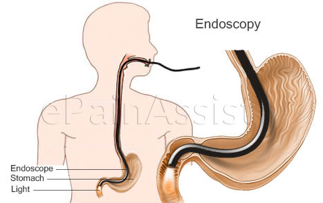
Endoscopy for Abdominal Pain or Stomach Ache

  • Laparoscopy
  • Endoscopy

Female Reproductive System and Pregnancy:

  • Pelvis: Exploration of pelvic abscess, laparoscopy.
  • Uterus: Dilation and Curettage – D and C.
  • Hysterectomy: Abdominal.
  • Hysterectomy: Vaginal, repair of prolapse uterus.
  • Endometriosis: Microwave Endometrial Ablation.
  • Ovaries: Oophorectomy.
  • Ectopic Pregnancy: Salpingo – oophorectomy – Ectopic Pregnancy.
  • Cancer of Cervix and Vagina: Excision of uterus, cervix, and vagina.

General Surgery:

  • Excision of Tumor: Colectomy, resection of rectum.
  • Gallbladder: Cholecystectomy.
  • Hernia: Repair.
  • Spleen: Splenectomy.
  • Ulcer: Excision.
  • Intestinal Obstruction: Laparotomy and bowel resection.
  • Volvulus and Intussusception: Laparotomy and bowel resection.

Kidney and Urinary System:

  • Bladder: Cystoscopy, Diathermy.
  • Kidney: Nephrectomy (Kidney Removal).
  • Nephrolithotomy: Removal of Kidney Stone, Retrograde Pyelogram.
  • Ureteral Stone: Ureterolithotomy.
  • Urethral Stone: Urethroscopy.

Male Reproductive System and Urinary Tract:

  • Urinary Retention: Prostatectomy.

References:

Also Read:

Physical Examination for Abdominal Pain or Stomach Ache: Inspection, Palpation, Auscultation

Physical Examination for Abdominal Pain or Stomach Ache Include Inspection, Palpation and Auscultation

Inspection Of The Abdomen:

Inspection is an observation of abdomen under optimum visualization. Inspection is to evaluate changes in abdominal contour, color and observation of any abnormal vascular changes.

  1. Abdominal Contour: Abdominal shape is often altered with certain disease. Inspection of abdominal curve may show a change of generalized distension or localized bulge.
    • Generalized Distension: Ascites (rounded symmetrical and bulge flank), hematoma – intra abdominal bleeding obesity, bowel distention by gas or liquid.
    • Localized: Portion of the abdomen shows swelling or bulge as in following diseases:
      • Right upper abdomen: Liver tumor, hepatitis.
      • Left upper abdomen: Splenomegaly, gastric tumor.
      • Upper half of the abdomen: Pancreatic cyst or tumor or acute gastric dilatation.
      • Lower half of the abdomen: Pregnancy, ovarian tumor, uterine fibroids, or bladder distention.
      • Scaphoid abdomen: Malnutrition.
    • Flank: Bulge in area of flank is secondary to disease of kidney tumor and kidney abscess.[1]
  2. Discoloration: Bluish discoloration seen with abdominal pain in following diseases:

    Umbilicus (Cullen's sign) and Flanks (Grey Turner's sign) can cause Abdominal Pain or Stomach Ache

    • Umbilicus (Cullen’s sign) – Bluish bruises and edema around umbilicus seen in acute pancreatitis and ectopic pregnancy.[2]
    • Flanks (Grey Turner’s sign)- Bluish bruising of flank seen on pancreatic necrosis, retroperitoneal bleeding and intra-abdominal bleeding.[3]
  3. Striae also known as stretch marks, striae atrophicae, vergetures, striae distensae, striae cutis distensae, striae gravidarum, lineae atrophica and linea albicante. Striae are a scarring of skin by separation of dermal elastic and fibrous tissues, commonly seen after pregnancy and loss of weight in obese patients. Abdomen is stretched considerably in obesity and pregnancy. After pregnancy and loss of weight in obesity stretch marks are very noticeable and detected in abdominal wall
  4. Surgical Scars: In most of the cases, scar is seen as a normal healing scar. In few cases, it may be presented as keloid or infected scar tissue.
  5. Abnormal Abdominal Wall Venous Flow: Changes in direction of venous flow is observed in patients experiencing partial or complete blockade of inferior vena cava by thrombosis or embolism. In a normal individual, blood in venous system of abdominal wall flows from umbilicus outward towards heart in upper half of abdomen and toward feet in lower half of abdominal wall. Blood flow changes direction in inferior vena caval blockage.
  6. Spider nevus of the abdominal wall: Also known as spider angioma and vascular spider. It is presented with central red bright spot of dilated arterioles and radiating branches spreading outward as spider web of venous branches carrying blood away from central arterioles. They are benign and seen in liver disease.
    • Hormonal Contraceptive: Higher estrogen level pregnant woman.
    • Liver Hepatic Disease: Unable to metabolize estrogen, cirrhosis.[4]
  7. Hernia: Bulge is observed in umbilical, epigastric, incisional and spigelian hernia.

Hernia Due To Abdominal Pain or Stomach Ache

Palpation Of The Abdomen:

After inspection, the patient is examined with palm and fingers of one or both hands. Palpation is either gentle superficial and deep or vigorous superficial and deep.

  1. Intra-abdominal Crepitus: Palpation may produce a sound and feel of crepitus. Crepitus is a crackling and popping sound. Intra-abdominal crepitus is observed in necrotizing enterocolitis occurs in premature infants.
  2. Abdominal Tenderness: Tenderness is defined as pain and discomfort when affected area is touched. Generalized tenderness – Peritonitis causes severe generalized abdominal pain and presents other symptoms such as loss of appetite, fever, dehydration, anorexia, poorly localized pain, rigidity, and rebound tenderness.

    Localized:

    • Appendicitis presented with localized tenderness in lower right quadrant of abdomen. Palpation over inflammatory mass such as appendix in right lower quadrant produces severe pain and tenderness.
    • Stomach cancer mass felt in left upper quadrant.[5]
  3. Mass: Diagnosis depends on location where mass is felt. Occasional cases liver, spleen and kidney may be palpable in normal individuals.

    Location:

    • Right Upper Quadrant: Liver hypertrophy, hepatic tumor, carcinoma of head of pancreases, gall bladder enlargement following cholecystitis.
    • Epigastric Mass: Gastric enlargement, pancreatic pseudocyst, pancreatic cancer or abdominal aneurysm.
    • Left Upper Quadrant: Splenomegaly.
    • Flank: Renal cysts, polycystic kidneys, and renal malignancies may all be visible in asthenic patients.
    • Lower Quadrants: Inflammatory or neoplastic disorders of the intestine. In the right lower quadrant appendicular abscess and cecal carcinoma are most likely, while in the left lower quadrant diverticular abscess or carcinoma of the sigmoid colon is most likely.
    • Hypogastric Quadrant: Pain cause by pelvic organs.

    Consistency:

    • Soft: Ovarian cyst, volvulus, bladder distention, cholecystitis.
    • Firm: Splenomegaly, neuroblastoma, kidney cancer, aortic aneurysm, bowel obstruction.
    • Hard: Uterine leiomyoma, stomach ulcer, hepatomegaly, liver cancer, kidney cancer, colon cancer, gallbladder tumor.
  4. Guarding: Peritonitis, appendicitis, diverticulitis, rupture ovarian cyst, twisted ovarian cyst, rupture aortic aneurysm, spleen rupture, uterus rupture, mesenteric embolism, pancreatitis, intestinal obstruction, ectopic pregnancy, rupture gallbladder, perforated ulcer, volvulus, intussusception, abdominal injury.
  5. Rigidity: Peritonitis, appendicitis, diverticulitis, rupture ovarian cyst, twisted ovarian cyst, rupture aortic aneurysm, spleen rupture, uterus rupture, mesenteric embolism, pancreatitis, intestinal obstruction, ectopic pregnancy, rupture gallbladder, perforated ulcer, volvulus, intussusception and abdominal injury.
  6. Rebound Tenderness (Rovsing’s sign): Tenderness and rebound tenderness on palpation. This is seen in appendicitis, peritonitis and ulcerative colitis.[6]

Auscultation Of The Abdomen:

Physical Examination for Abdominal Pain or Stomach Ache: Auscultation Of The Abdomen

  • Normal Peristalsis: Normal peristalsis of the intestine produces bowel sounds as gas and fluid are passed through the intestinal lumen. Normally, the bowel sounds are intermittent, low-pitched, chuckling sounds. Bowel sounds may be decreased or increased in few diseases.
  • Absence of Bowel Sound (paralytic ileus): Late stage of intestinal obstruction, paralytic ileus seen in congestive heart failure, pneumonia and uremia.
  • Hyperactive Peristalsis(rushes): High pitch and tinkling sound, earlier phase of intestinal obstruction.
  • Rubs: Irritation of inflamed serosa surface of peritoneum rubbing against diseased organs during inspiration and expiration of breathing cycle e.g. liver, spleen and abdominal mass. Normal peritoneal surface when inflamed from infection or neoplasm causes adherence to vascular structure and surrounding organ.
  • Vascular Bruits: Audible turbulent blood flow conducting swishing sound over major arteries during systole. Observed in aortic aneurysm and atherosclerosis of aorta and renal artery. Occasional bruits are heard during systole and diastole in a patient suffering with arteriovenous communications.
  • Crepitus: Sound is produced by friction between gas and fluid within the viscous of organ or air entrapped in subcutaneous tissue following infection such as gas gangrene. Penetrating chest wall injury can cause subcutaneous emphysema secondary to air leaking from alveoli into subcutaneous tissue through a track of air fistula. Post surgery claustredial infection can cause intra-abdominal gas gangrene.

Medical Emergency:

Physical Examination for Abdominal Pain or Stomach Ache: Medical Emergency

  • Vomiting: Persistent and continuous vomiting causes dehydration.
  • Diarrhea: Continuous several episodes causing dehydration.
  • Profuse hemoptysis: Peptic perforation, gastric bleeding tumor. Bleeding per rectum- hemorrhoids, tumor.
  • Bleeding Abdomen: Ruptured aneurysm.
  • Peritonitis caused by Perforation of Abdominal Organs. Ulcer from esophagus, stomach, intestine and large bowel can perforate and cause peritonitis. Gastric carcinoma, intestinal tumor, inflamed appendix, Meckel’s diverticulitis or intestinal infarction can cause perforation and severe peritonitis. Rare causes of peritonitis are ingestion of foreign bodies and abdominal surgery. Auto or work injury may cause blunt or penetrating abdominal trauma causing injury to internal organ and tear. Peritonitis is cause by feces in the peritoneal cavity inducing E.Coli and anaerobic bacterial (bacteroides fragilis) infection. Surgical trauma or injury causes peritonitis mostly infected by staphylococcus aureus or coagulase negative staphylococci and may include fungi and Candida.
  • Non-infected Peritonitis: Sterile fluid from body if leaked into peritoneal cavity causes inflammatory reaction. Non-infected peritonitis is observed with leakage of blood, endometriosis, gastric juice, urine, menstruum, pancreatic juice or ovarian cyst rupture.[7]

References:

Also Read: