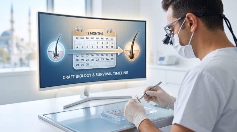
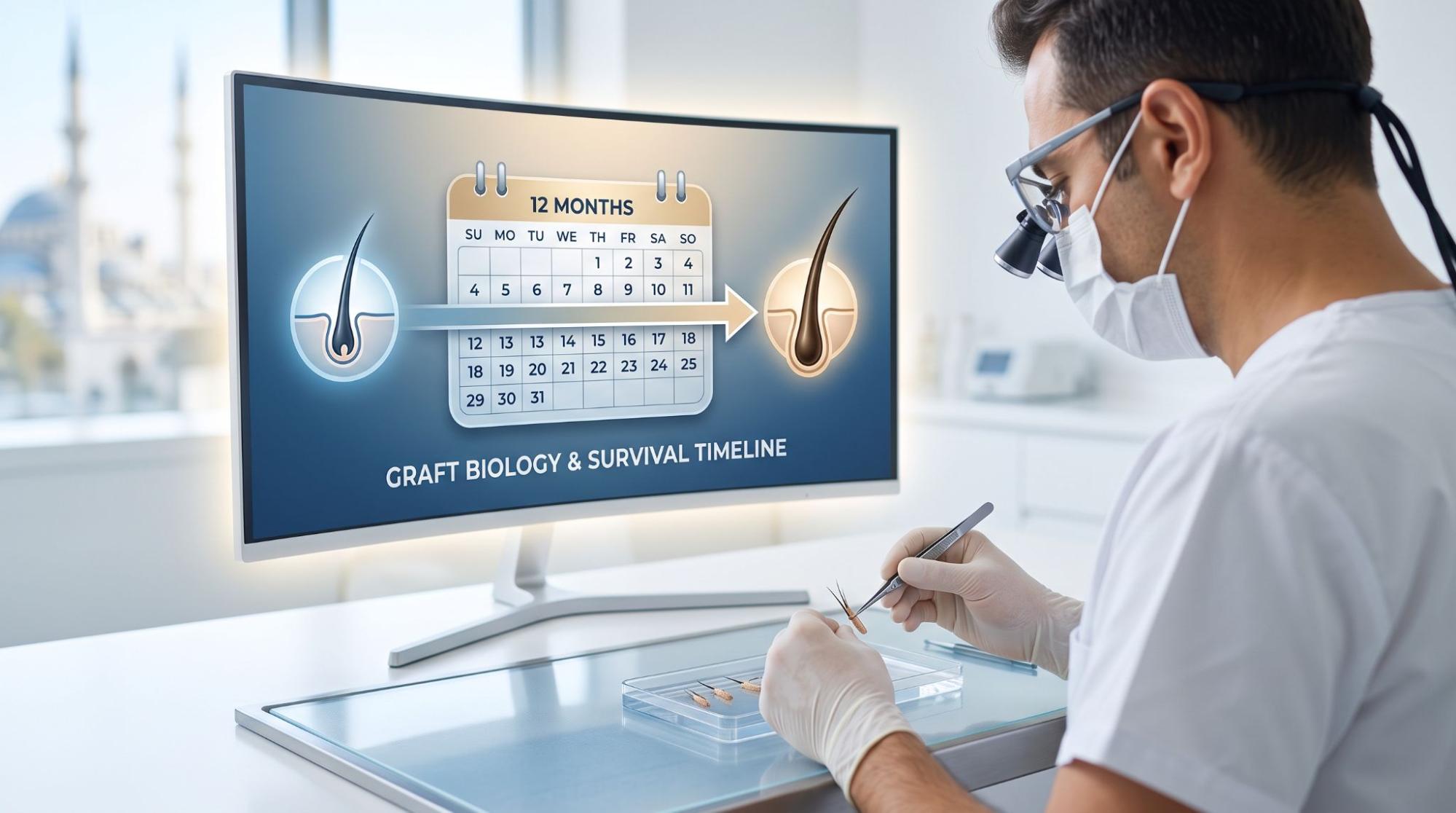
The Biological Stages of Hair Graft Survival: Understanding the Post-Surgical Healing Spectrum

0

For millions of men and women experiencing androgenetic alopecia, modern surgical hair restoration—utilizing Follicular Unit Extraction (FUE) or Direct Hair Implantation (DHI)—provides a permanent, natural-looking solution. However, achieving superior cosmetic density depends on much more than the surgeon’s skill during extraction and incision creation. The long-term success of any hair transplant is deeply rooted in human follicular biology and the complex physiological stages of graft survival.

Understanding the cellular journey of transplanted follicles allows prospective patients to set realistic expectations, maintain proper post-operative compliance, and navigate the recovery journey with confidence.

The Cellular Biology of Graft Integration: Ischemia and Neovascularization

When a hair follicle is harvested from the androgen-resistant donor zone (typically the occipital region at the back of the scalp), it is temporarily severed from its primary blood supply. Once re-implanted into the recipient site, a critical biological race against ischemic tissue damage begins.

The Biological Stages of Hair Graft Survival: Understanding the Post-Surgical Healing Spectrum

During the initial 72 hours post-surgery, transplanted grafts survive through a mechanism known as plasmatic imbibition—absorbing essential nutrients and oxygen purely from surrounding interstitial tissue fluid. Within 48 to 7 days, micro-vascular connections (neovascularization) begin to re-establish, connecting the follicle back to the body’s systemic blood circulation.

According to clinical research published by the National Institutes of Health (NCBI), transplanted hair follicles naturally enter a protective resting phase following surgical trauma, causing the visible hair shaft to shed while the underlying dermal papilla remains fully viable beneath the skin.

This temporary shedding phase—often referred to as “shock loss”—typically occurs between weeks 2 and 6 post-operation. While it can cause temporary concern for patients, it is a completely normal physiological reaction. The follicle root remains intact, preparing its metabolic machinery to initiate a new, permanent anagen (growth) phase.

Chronological Growth Progression: What to Expect Month by Month

Patience is a fundamental requirement in hair restoration surgery. Because human hair grows at an average rate of 1 to 1.5 centimeters per month, visual changes occur gradually over an extended timeframe rather than overnight.

The standard visual recovery timeline generally unfolds across four key biological stages:

  • Months 1 to 3 (The Dormant Phase): Post-surgical redness and minor inflammation subside. The majority of transplanted shafts shed, and the follicles establish deep vascular anchors within the dermal layer.
  • Months 4 to 6 (Initial Emergence): Fine, unpigmented hairs begin to breach the scalp surface. Early growth may appear sporadic, fine, or slightly texturally altered as the new shaft matures.
  • Months 7 to 9 (Density Amplification): Hair shafts undergo rapid thickening (caliber expansion), gain natural pigmentation, and provide noticeable visual coverage across the hairline or crown.
  • Months 10 to 12 (Maturation & Final Density): The vast majority of patients achieve their final visual result by the one-year mark, featuring full cosmetic density and seamless integration with existing native hair.

To follow the detailed biological evolution month by month and track key recovery benchmarks, reviewing the TurkHairIndex step-by-step post-operative recovery guide offers essential insights into what patients should expect at every phase of the healing journey.

Key Biological Factors Influencing Graft Yield

While patient physiology plays a significant role in healing, several clinical and post-operative factors directly impact final graft survival rates:

  1. Out-of-Body Time & Holding Solutions: Minimizing the time grafts spend outside the body and keeping them hydrated in chilled holding solutions (such as HypoThermosol or saline) prevents cellular apoptosis.
  2. Scalp Hygiene & Scab Management: Gentle washing protocols starting on day 3 help eliminate crusting and minimize the risk of localized folliculitis without dislodging fragile grafts.
  3. Vascular Health & Lifestyle Compliance: Avoiding nicotine, strenuous physical exercise, and direct sun exposure during the first 14 days preserves micro-vascular blood flow to the newly implanted roots.

Conclusion

Hair restoration is a harmonious blend of surgical artistry and human dermatological physiology. By understanding the underlying cellular cycle of hair grafts and adhering strictly to post-operative guidelines, patients can navigate their recovery timeline with peace of mind and achieve lasting, natural-looking hair density.

Does a Bucket-Handle Meniscus Tear Always Need Surgery? Repair vs Meniscectomy and Recovery

A diagnosis of a bucket-handle meniscus tear often leads to an immediate question: Does this mean I need surgery?

The answer is not quite as simple as yes or no.

Some meniscus tears can be treated without surgery, particularly when they are small, stable, and do not interfere with normal knee movement. A bucket-handle tear, however, is a different type of injury. In this tear pattern, a long portion of the meniscus separates and may flip inward toward the center of the knee. When that fragment becomes displaced, it can catch inside the joint and sometimes prevent the knee from straightening completely.

That is why bucket-handle tears are more likely than many other meniscus injuries to require arthroscopic surgery.

Even when surgery is recommended, another important decision remains: Should the torn meniscus be repaired, or should the damaged portion be removed with a partial meniscectomy?

Whenever the tissue can realistically heal, preserving the meniscus through repair is generally preferred because the meniscus plays an important role in distributing force across the knee and protecting the joint cartilage. But not every bucket-handle tear can be successfully repaired.

The decision depends on where the tear is located, how healthy the meniscus is, how long the tear has been present, whether the displaced tissue can be repositioned, the condition of the rest of the knee, and several other factors. Current guidance emphasizes preserving as much functional meniscal tissue as possible when surgery is necessary.

What Is a Bucket-Handle Meniscus Tear?

Each knee contains two menisci: the medial meniscus on the inner side of the knee and the lateral meniscus on the outer side. These crescent-shaped structures sit between the thighbone and shinbone and help distribute weight, absorb shock, stabilize the knee, and protect the articular cartilage.

A bucket-handle tear is usually a large vertical or longitudinal meniscus tear. One part of the torn meniscus remains attached at both ends while the central portion can move inward.

The appearance is sometimes compared with the handle of a bucket, which is where the injury gets its name.

If the torn fragment stays close to its normal position, symptoms may be less dramatic. When it flips into the middle of the knee joint, however, it can physically obstruct movement.

Common symptoms include pain along the joint line, swelling, clicking or catching, difficulty bending or straightening the knee, a feeling that something is moving inside the joint, and episodes in which the knee becomes locked.

Magnetic resonance imaging is commonly used to confirm the tear and evaluate its location, size, displacement, tissue quality, and associated injuries. It is currently the preferred imaging method for diagnosing acute meniscal tears.

Does a Bucket-Handle Meniscus Tear Always Need Surgery?

No, not every bucket-handle meniscus tear automatically requires surgery. However, a true displaced bucket-handle tear is one of the meniscus tear patterns in which surgery is relatively common.

The distinction between a stable tear and a displaced tear is important.

If imaging shows a bucket-handle-type tear but the fragment remains relatively stable, the knee has a full range of motion, symptoms are manageable, and there is no significant catching or locking, a doctor may occasionally consider a period of conservative treatment.

That can include activity modification, temporary avoidance of twisting and deep squatting, physical therapy, swelling control, and gradual strengthening.

Nonoperative treatment can be appropriate for selected acute meniscus injuries. Evidence specifically addressing nonsurgical treatment of traumatic bucket-handle tears remains limited, however, and international consensus recommendations note that bucket-handle tears—particularly larger or mechanically significant tears—may require earlier surgical intervention.

The situation changes significantly when the torn fragment has moved into the joint and the knee cannot straighten normally.

A Locked Knee Makes Surgery More Likely

A locked knee from a bucket-handle meniscus tear is different from a knee that simply feels stiff because of pain or swelling.

True mechanical locking occurs when the displaced meniscal fragment physically blocks normal knee motion. A person may be unable to fully straighten the leg even when trying to relax the muscles.

A displaced acute meniscus tear that restricts knee movement is generally considered a reason for relatively prompt orthopedic assessment and often surgical treatment. Current clinical guidance specifically recognizes displaced or displacing meniscus tears that restrict range of motion as injuries that can benefit from early surgery.

This does not necessarily mean that every patient needs emergency surgery the same day. It does mean that repeatedly trying to exercise through a mechanically locked knee or waiting months without orthopedic assessment may not be appropriate.

Can a Bucket-Handle Meniscus Tear Heal Without Surgery?

People frequently search for ways to make a bucket-handle meniscus tear heal naturally, but the answer depends heavily on the tear’s location and stability.

The meniscus does not receive the same blood supply throughout its entire width.

The outer region, often called the red-red zone, has the best blood supply and therefore the greatest healing potential. The transitional red-white zone has a more limited blood supply. The inner white-white zone receives very little blood and is much less capable of healing.

A stable tear near the vascular outer portion has more potential to heal than a displaced tear extending into poorly vascularized tissue.

There is another problem with a displaced bucket-handle tear: healing requires the torn surfaces to remain positioned appropriately. If a large fragment is folded into the center of the knee, the tissue may not simply reconnect on its own.

For this reason, the question is not only whether meniscal tissue can heal. The doctor also has to determine whether the displaced fragment can remain reduced in its normal position long enough for healing to occur.

Why Doctors Try to Repair a Bucket-Handle Meniscus Tear When Possible

Years ago, removal of damaged meniscal tissue was much more common. Today, there is greater emphasis on meniscus preservation.

The reason is mechanical as much as biological.

The meniscus spreads loads across the knee. Removing part of it decreases the amount of tissue available to distribute those forces. The remaining joint surfaces therefore experience greater contact stress.

Over many years, loss of meniscal tissue is associated with an increased risk of degenerative changes in the knee. Long-term studies following partial meniscectomy have repeatedly demonstrated more radiographic osteoarthritis in operated knees than in comparison knees.

For a bucket-handle tear, this concern can be particularly important because the torn segment may represent a substantial portion of the meniscus.

If that entire segment can be repaired rather than removed, the knee retains considerably more of its natural shock-absorbing tissue.

That is why the goal is increasingly save the meniscus when the meniscus is worth saving, rather than automatically removing every torn portion.

Bucket-Handle Meniscus Repair: What Happens During Surgery?

A bucket-handle meniscus repair is usually performed arthroscopically through several small openings around the knee.

The surgeon first examines the tear directly and determines whether the displaced fragment can be returned to its normal position.

This is called reduction of the tear.

The surgeon then evaluates tissue quality. A meniscus that looks healthy and can hold sutures offers a better chance of successful repair than severely frayed, degenerative, crushed, or chronically deformed tissue.

If the tear is repairable, sutures or specialized meniscal fixation devices are placed across the tear so the two sides remain together while biological healing occurs.

Several repair techniques are available, including all-inside, inside-out, and outside-in techniques. The technique selected depends partly on the tear’s location, size, surgeon preference, and anatomy.

Who Is a Good Candidate for Bucket-Handle Meniscus Repair?

Age matters, but it is not the only consideration.

A relatively young person with a traumatic tear, healthy cartilage, good meniscal tissue, and a tear extending through an area with reasonable blood supply is often an attractive candidate for repair.

But being over a certain age does not automatically make repair impossible.

A physically active adult with good meniscal tissue and little arthritis may still have a repairable tear, while a much younger patient with badly damaged tissue may not.

The surgeon generally considers the location of the tear, quality of the meniscus, blood supply, ability to reduce the displaced fragment, length of the tear, time since injury, amount of existing arthritis, associated ligament injury, and overall stability of the knee.

The final decision sometimes cannot be made with complete certainty from magnetic resonance imaging alone. The surgeon may need to inspect and probe the tissue during arthroscopy before deciding whether a durable repair is realistic.

When Is Partial Meniscectomy Used for a Bucket-Handle Tear?

A partial meniscectomy does not usually mean that the entire meniscus is removed.

Instead, the surgeon trims away tissue that cannot be repaired while preserving as much healthy and stable meniscus as possible.

Partial meniscectomy may be considered when the bucket-handle fragment is badly damaged, the tissue is severely degenerative, the tear lies predominantly in an area with poor healing potential, the fragment cannot be securely reduced, or a previous repair has failed and another repair is unlikely to succeed.

Poor-quality loose meniscal tissue causing catching and pain is one situation in which trimming may be appropriate.

This distinction is important because patients sometimes hear that meniscectomy provides a quicker recovery and assume it must therefore be the better procedure.

It is usually more accurate to think of partial meniscectomy as the appropriate option when the torn tissue is not realistically salvageable, rather than as the preferred shortcut to faster recovery.

Meniscus Repair vs Partial Meniscectomy: Which Is Better?

There is no single answer that applies to every bucket-handle tear.

If a tear has good healing potential, meniscus repair offers one major advantage: more of the natural meniscus remains in the knee.

The tradeoff is that the repaired tissue must heal.

That means rehabilitation is longer, activity restrictions are greater, and there is a possibility that the repair will fail or the meniscus will tear again.

Partial meniscectomy has the opposite profile.

There is no repaired tear that must biologically unite, so patients can often put weight on the leg sooner, regain motion faster, and return to everyday activities more quickly.

The downside is permanent loss of part of the meniscus.

Research comparing the procedures has consistently highlighted this tradeoff. Meniscus repair tends to have a higher chance of requiring another operation, whereas preservation of the meniscus is associated with better long-term joint preservation in appropriately selected patients.

For that reason, modern treatment does not simply ask, “Which operation has the easiest recovery?”

A better question is: Can this meniscus be saved successfully enough to justify the longer recovery?

How Successful Is Bucket-Handle Meniscus Repair?

Bucket-handle tears can be challenging repairs because they are usually large and the displaced tissue may have been subjected to repeated stress.

Still, many repairs heal successfully.

A systematic review and meta-analysis specifically evaluating bucket-handle repairs estimated an overall failure rate of approximately 14.8 percent, although individual studies varied considerably. Medial meniscus repairs and isolated repairs showed higher failure rates in that analysis.

Longer-term studies illustrate why the decision can be complicated. Some cohorts have found higher reoperation or failure rates over many years, while successful repairs have also been associated with better knee function and fewer degenerative changes than failed repairs.

A more recent study following patients for an average of about seven years reported good clinical outcomes and a repair failure rate of approximately 9 percent, demonstrating that large bucket-handle tears can still have durable results when appropriately selected and repaired.

These numbers should not be interpreted as predicting what will happen to an individual patient. Repair success varies significantly according to tear characteristics, tissue quality, associated injuries, rehabilitation, and how failure is defined in a particular study.

What Happens If a Bucket-Handle Meniscus Tear Is Left Untreated?

Not every untreated bucket-handle tear deteriorates at the same rate.

However, a persistently displaced tear can continue producing pain, swelling, catching, and loss of motion. The mobile fragment may repeatedly move within the joint, and the person may begin avoiding normal knee movement.

Long-standing restriction can also contribute to weakness and stiffness.

Delay may become particularly relevant when the goal is meniscus repair. A newly displaced, healthy fragment may be easier to reduce and repair than tissue that has remained displaced long enough to become deformed, scarred, or damaged.

Current guidance therefore suggests considering earlier intervention when a symptomatic acute tear is likely to be repairable, particularly when displacement limits motion.

Someone whose knee is stuck and cannot fully straighten should therefore seek orthopedic evaluation rather than assuming the problem can be stretched out over time.

Bucket-Handle Meniscus Repair Recovery

The biggest disadvantage of meniscus repair from the patient’s perspective is often the rehabilitation period.

A repair must be protected long enough for the tissue to heal.

Exactly how much weight can be placed on the leg and how far the knee can bend depends on the location, size, stability, and surgical technique used for the repair. Rehabilitation protocols therefore vary.

Some patients are allowed early weight bearing with the knee protected in a brace, while others need crutches and restricted weight bearing for a period of time. Knee bending is commonly progressed gradually during the first several weeks.

Early rehabilitation typically focuses on swelling control, restoring safe knee extension, gradually improving flexion, maintaining quadriceps activation, and walking safely.

Strengthening is progressively increased as healing advances.

Running, jumping, deep loaded knee flexion, rapid pivoting, and sport-specific cutting movements are introduced later because they place considerably greater stress across the healing meniscus.

A return to normal activity around six months is common after repair, although athletes involved in pivoting or high-demand sports may require six to nine months depending on healing, strength, symptoms, and functional testing. Recent international rehabilitation guidance recommends combining time after surgery with objective recovery milestones rather than allowing return to sport based only on the calendar.

Partial Meniscectomy Recovery Is Usually Faster

Recovery following partial meniscectomy for a bucket-handle tear is typically much quicker because there is no repaired tissue that needs to join together.

After an uncomplicated arthroscopic partial meniscectomy, early full weight bearing is commonly permitted as symptoms allow. Crutches may be used temporarily until the person can walk without a significant limp.

Swelling and quadriceps weakness still need to be addressed, and a rapid recovery does not mean rehabilitation can be skipped.

Patients usually progress through range-of-motion exercises, quadriceps strengthening, balance work, walking, and then higher-level activities.

General recovery estimates are around three to six weeks for partial meniscectomy, compared with approximately three to six months or longer after repair. Return to sport after meniscectomy is often within approximately four to twelve weeks, whereas six to nine months is commonly recommended after meniscus repair.

These are averages rather than deadlines. Persistent swelling, weakness, pain, instability, or restricted movement should take priority over a predetermined return date.

Why Faster Recovery Does Not Automatically Mean a Better Operation

It is understandable to prefer the procedure that allows quicker walking, working, and exercising.

But the purpose of meniscus surgery extends beyond the first few weeks after the operation.

Imagine that a repair requires several months of rehabilitation but preserves a large segment of healthy meniscus that could remain functional for many years. In the right patient, accepting that longer recovery may provide a worthwhile long-term benefit.

Conversely, attempting to repair severely damaged tissue simply because preservation sounds preferable can expose the patient to months of restrictions followed by a failed repair and another operation.

The ideal treatment therefore balances healing potential, meniscus preservation, short-term recovery, risk of another operation, and long-term knee health.

Can a Bucket-Handle Tear Come Back After Repair?

Yes.

A repaired meniscus can fail to heal completely or can tear again after initially healing.

This does not necessarily mean the original surgical decision was wrong. Meniscus repair deliberately accepts some risk of reoperation in exchange for an opportunity to preserve functional meniscal tissue.

If symptoms return after repair, particularly recurrent locking, joint-line pain, swelling, or catching, the surgeon may order another examination and sometimes repeat magnetic resonance imaging.

Depending on what is found, another repair may occasionally be possible. In other cases, the damaged section may ultimately require partial meniscectomy.

Does a Bucket-Handle Meniscus Tear With an Anterior Cruciate Ligament Injury Change Treatment?

Bucket-handle tears sometimes occur together with an anterior cruciate ligament tear, particularly after twisting sports injuries.

These cases require a somewhat different discussion because knee stability and meniscus healing are interconnected.

When anterior cruciate ligament reconstruction and meniscus repair are performed together, the surgeon may try to preserve the meniscus whenever practical. Some studies of bucket-handle tears have reported favorable outcomes when repairs are performed alongside anterior cruciate ligament reconstruction.

Treatment must still be individualized. The size of the bucket-handle tear, meniscus involved, cartilage condition, knee stability, activity level, and reconstructive plan all influence the final approach.

How Soon Should a Bucket-Handle Meniscus Tear Be Treated?

There is no universal rule saying every bucket-handle tear must be operated on within a particular number of days.

Symptoms matter.

A relatively stable tear in a knee that bends and straightens normally may allow more time for assessment and treatment planning.

A displaced fragment causing a genuinely locked knee deserves more prompt attention.

Early evaluation may also matter when the surgeon hopes to repair the meniscus because prolonged displacement can potentially make reduction or repair more difficult.

The practical message is that bucket-handle tear plus inability to fully straighten the knee should not be treated like ordinary knee soreness.

Does Everyone With a Bucket-Handle Meniscus Tear Need Magnetic Resonance Imaging?

Magnetic resonance imaging is usually extremely helpful because it shows the meniscus and other soft tissues that cannot be evaluated adequately on a standard X-ray.

It can identify the tear pattern, show whether the meniscus is displaced, and look for associated injuries involving structures such as the anterior cruciate ligament or cartilage.

Magnetic resonance imaging is currently considered the preferred imaging method for suspected acute meniscal tears because of its diagnostic accuracy.

However, imaging is only one part of the decision. Symptoms, physical examination findings, range of motion, age, activity demands, knee arthritis, and what the surgeon actually finds during arthroscopy can all affect treatment.

When Should You Seek Medical Attention Quickly?

A knee injury warrants prompt medical assessment when the knee becomes stuck and cannot fully straighten, swelling is substantial after an injury, weight bearing is very difficult, or repeated catching and locking continue.

After meniscus surgery, worsening rather than improving pain, persistent or recurrent major swelling, increasing stiffness, new neurological symptoms, symptoms suggesting infection, or signs concerning for a blood clot also require medical assessment.

So, Does a Bucket-Handle Meniscus Tear Need Repair or Meniscectomy?

For many patients, the most important question is not simply “Do I need surgery?”

It is “If I do need surgery, can my meniscus be saved?”

A stable bucket-handle tear without significant mechanical symptoms may occasionally be managed without an operation. A displaced bucket-handle tear that causes true locking or prevents normal knee motion is much more likely to require arthroscopic treatment.

When surgery is necessary and the tissue has reasonable healing potential, meniscus repair is generally favored because preserving the meniscus helps maintain normal load distribution within the knee.

Partial meniscectomy remains valuable when a tear is not realistically repairable. It usually provides a considerably faster recovery, but that advantage comes at the cost of permanently removing some meniscal tissue.

The best treatment therefore cannot be determined simply by the words “bucket-handle tear” on a magnetic resonance imaging report.

The surgeon needs to determine whether the fragment is displaced, whether it can be reduced, where the tear sits within the meniscus, whether the tissue can hold a repair, how much arthritis is already present, and what other injuries exist within the knee.

For a repairable tear, several months of rehabilitation may be worthwhile if it preserves functioning meniscus for the years ahead. For a badly damaged or nonhealing fragment, carefully removing only the unsalvageable portion may provide the more reliable result.

The goal is not simply to make the knee feel better quickly. Whenever possible, it is to restore movement while preserving as much useful meniscus as the knee can keep.

Burning Sensation on One Side of the Lower Back: Causes, Symptoms and Treatment

A burning sensation on just one side of the lower back can be difficult to interpret. It may feel as though a patch of skin is unusually hot, as if something is burning beneath the skin, or like a deep warmth coming from the muscles. For some people, the sensation stays in one spot. For others, it spreads toward the buttock, hip, thigh, or leg.

One-sided burning in the lower back is not a diagnosis by itself. The sensation can come from muscles, spinal nerves, the skin, joints around the pelvis, or occasionally an internal organ. The location alone also does not tell you exactly what is wrong. Burning on the left side and burning on the right side can have many of the same causes.

What often matters more is what happens along with the burning.

Is there numbness or tingling? Does the discomfort travel down the leg? Is the skin sensitive to touch? Did a rash appear? Does the area actually feel hot? Are there urinary symptoms or fever?

Those details can help distinguish a relatively simple muscle problem from nerve irritation, shingles, a kidney problem, or another condition that deserves medical attention.

What Does a Burning Sensation on One Side of the Lower Back Feel Like?

People use the word “burning” to describe several different sensations.

You may notice:

  • A hot patch on the left or right lower back
  • Burning beneath the skin
  • A warm sensation even though the skin feels normal
  • Tingling or pins and needles
  • Stinging
  • Prickling
  • Electric shock-like pain
  • Sharp pain mixed with burning
  • Skin sensitivity
  • Numbness
  • Burning that travels into the buttock
  • Burning that runs down one leg

Burning, electrical sensations, tingling, numbness, and pain triggered by light touch are commonly associated with neuropathic pain, although these descriptions alone cannot prove that a nerve is the cause.

The pattern is therefore important. A burning spot after lifting a heavy object may have a very different explanation from burning that shoots from the lower back into the foot.

Why Is the Burning Only on One Side of the Lower Back?

Many structures in the lower back exist as pairs or affect one side more than the other.

A muscle can be strained predominantly on one side. One spinal nerve root may become irritated. One sacroiliac joint can become painful. Shingles commonly affects a band of skin on one side of the body. Kidney conditions may also produce pain predominantly on one side.

This is why unilateral lower back burning can arise from several unrelated problems.

It is also important not to assume that the left side means one condition and the right side means another. In most musculoskeletal and nerve-related problems, either side can be affected.

Common Causes of Burning on One Side of the Lower Back

1. Irritated or Compressed Nerve in the Lower Spine

A nerve problem is one of the first possibilities to consider when the sensation is distinctly burning, tingling, or electric.

The spinal nerves leave the lower spine and travel into the pelvis and legs. If one nerve root becomes irritated or compressed, symptoms may develop primarily on that side.

Possible symptoms include:

  • Burning lower back pain
  • Tingling
  • Pins and needles
  • Numbness
  • Shooting pain
  • Electric shock-like pain
  • Pain extending into the buttock
  • Leg pain
  • Weakness in part of the leg or foot

A herniated disc, age-related changes in the spine, or narrowing around a nerve can all contribute to nerve irritation. Structural causes of back pain include herniated discs, spinal stenosis, muscle strain, ligament sprain, and other conditions that may irritate nearby nerves.

Burning that remains only in the lower back does not automatically mean a nerve is involved, but burning combined with tingling, numbness, or radiating pain makes nerve irritation more likely.

2. Sciatica

Sciatica is a particularly common explanation when burning begins in the lower back or buttock and then travels down one leg.

The pain may feel:

  • Burning
  • Sharp
  • Shooting
  • Electric
  • Aching

Some people also develop numbness, tingling, or weakness.

Sciatica usually affects one side of the body and often follows a path from the lower back into the buttock and down the back of the thigh or calf. Sitting for a long time, coughing, or sneezing may worsen symptoms in some people.

A burning sensation that stays only in a small patch of the lower back is less typical of classic sciatica. However, if the discomfort travels down the buttock or leg, sciatica becomes a stronger possibility.

3. Herniated Disc

A spinal disc sits between adjacent vertebrae and helps absorb pressure.

When part of a disc pushes outward, it may irritate a nearby spinal nerve. Because the nerve on one side may be affected more than the other, the symptoms can be distinctly one-sided.

A herniated disc may cause:

  • Burning lower back pain
  • Pain in one buttock
  • Shooting leg pain
  • Tingling
  • Numbness
  • Weakness

Not every herniated disc causes symptoms. The problem becomes more noticeable when the disc irritates or compresses surrounding nerves.

4. Muscle Strain on One Side of the Lower Back

Burning does not always mean nerve damage.

A muscle strain can occur predominantly on one side, particularly after twisting, lifting, reaching, exercising, or repeatedly using one side of the body.

You may notice:

  • Localized soreness
  • Tightness
  • Tenderness
  • Burning after activity
  • Muscle spasm
  • Pain while bending or twisting
  • Pain when getting up from a chair or bed

Muscle and tendon strains are among the common mechanical causes of lower back pain.

A muscular cause becomes more likely when the symptoms began shortly after an unusual activity and there is no numbness, tingling, leg weakness, rash, or fever.

For example, spending several hours gardening, moving furniture, lifting boxes, or performing an unfamiliar exercise can overload one side of the lower back.

5. Myofascial Pain

Muscles and the connective tissue surrounding them can develop painful, tight areas that produce localized discomfort.

Myofascial pain may feel:

  • Aching
  • Tight
  • Tender
  • Deep
  • Occasionally burning

Certain spots may be especially sensitive when pressed.

Myofascial pain involving the muscles that support the spine is another recognized cause of lower back discomfort.

Unlike sciatica, myofascial pain usually does not follow a clear nerve pathway down the leg.

Burning Sensation on One Side of the Lower Back Without a Rash

A particularly common question is whether burning on one side of the back without a rash could still be shingles.

It can.

Shingles may begin with:

  • Burning
  • Pain
  • Tingling
  • Itching
  • Skin sensitivity

These symptoms can start several days before the rash becomes visible.

The rash typically develops as a strip or cluster of blisters on one side of the body and usually does not cross the middle of the body.

Therefore, unexplained one-sided skin burning should be watched carefully, particularly if the area becomes unusually sensitive to clothing or light touch.

Not every case of burning without a rash is shingles. Nerve irritation from the spine can create very similar sensations.

6. Shingles

Shingles is especially important when burning affects a relatively narrow area on one side of the back.

Before the rash appears, the person may feel as though the skin is:

  • Burning
  • Tingling
  • Itching
  • Stinging
  • Painful to touch

The rash then usually appears in a band on one side of the trunk.

This pattern occurs because shingles affects sensory nerves supplying a particular region of skin.

Some people develop marked sensitivity where ordinary contact from a shirt, bedsheet, or hand feels painful.

If a painful or blistering rash develops after several days of unexplained burning, medical evaluation should be obtained promptly.

7. Sacroiliac Joint Pain

The sacroiliac joints connect the lower spine to the pelvis. There is one joint on each side.

Irritation or inflammation in one sacroiliac joint can cause pain predominantly on the left or right side of the lower back.

The pain may extend into:

  • The buttock
  • Hip
  • Upper thigh

Symptoms can become worse after remaining in one position for a long time or during movements that rotate the pelvis.

Sacroiliac joint pain is more often described as aching, sharp, or deep rather than purely burning. However, some people may use “burning” to describe their discomfort, particularly when surrounding tissues or nerves are irritated.

A healthcare professional may need to distinguish sacroiliac joint pain from lumbar nerve pain because the symptoms can overlap.

Burning on the Left Side of the Lower Back

Burning on the left side can come from the same spinal, muscular, nerve, skin, or joint problems that affect the right side.

Common possibilities include:

  • Left-sided muscle strain
  • Left lumbar nerve irritation
  • Left-sided sciatica
  • Herniated disc affecting a left nerve root
  • Left sacroiliac joint pain
  • Shingles
  • Kidney problems

The side alone rarely establishes the diagnosis.

Instead, look for the pattern.

If the burning extends into the left buttock and leg with tingling or numbness, nerve irritation is more likely.

If the skin becomes hypersensitive and a rash develops, shingles becomes more likely.

If the discomfort is deeper in the side or flank and occurs with urinary symptoms, the urinary system needs consideration.

Burning on the Right Side of the Lower Back

The same principle applies to right-sided burning.

Possible causes include:

  • Right-sided muscle strain
  • Right-sided sciatica
  • Lumbar radiculopathy
  • Herniated disc
  • Sacroiliac joint irritation
  • Shingles
  • Kidney stone or kidney infection

Pain that began after lifting or twisting is more suggestive of a musculoskeletal cause, especially when movement reproduces the discomfort.

Burning or electric sensations that travel into the right leg raise more concern for nerve irritation.

Deep side pain with fever or urinary changes should not be assumed to be coming from the spine.

8. Kidney Infection

Kidney problems are often blamed for lower back discomfort, but kidney pain tends to be located more toward the side or flank and may be somewhat higher than ordinary lumbar muscle pain.

A kidney infection may produce:

  • Back or side pain
  • Fever
  • Chills
  • Pain or burning during urination
  • Frequent urination
  • Cloudy urine
  • Bloody urine
  • Foul-smelling urine
  • Nausea
  • Vomiting

An isolated burning patch over the skin of the lower back without fever or urinary symptoms is less typical of a kidney infection.

If one-sided back pain occurs together with fever, chills, or urinary symptoms, medical assessment should not be delayed.

9. Kidney Stones

Kidney stones can also cause severe pain on one side of the back or flank.

The pain may come in waves and can spread toward:

  • The lower abdomen
  • Groin
  • Pelvic region

Other symptoms can include:

  • Blood in the urine
  • Frequent urge to urinate
  • Pain during urination
  • Nausea
  • Vomiting
  • Cloudy or foul-smelling urine

Kidney stone pain is typically deeper and often much more intense than a superficial burning sensation in the skin.

What Does It Mean If the Skin Feels Hot but Looks Normal?

Sometimes a person is convinced that one side of the lower back is hot, yet another person touches the area and notices no temperature difference.

This can happen with altered sensory nerve signals.

Neuropathic pain may be experienced as burning, electrical, tingling, or temperature-related discomfort even without visible skin changes.

In this situation, pay attention to whether the burning is accompanied by:

  • Tingling
  • Numbness
  • Electric pain
  • Abnormal sensitivity
  • Pain from light touch
  • Radiation into the leg

These features strengthen the possibility of nerve involvement.

What If One Side of the Lower Back Is Actually Hot to Touch?

True skin warmth is different from the sensation of internal burning.

If one side of the back is objectively hotter, check for:

  • Redness
  • Swelling
  • Rash
  • Blisters
  • Tenderness
  • Broken skin
  • Drainage

An inflammatory or skin-related condition may be present.

A hot, red, swollen area should not automatically be attributed to a pinched nerve.

If warmth is accompanied by fever, rapidly increasing redness, or significant tenderness, medical evaluation is appropriate.

Burning Lower Back Pain That Goes Into the Buttock

Burning that spreads from one side of the lower back into the buttock can occur with several conditions.

Possible causes include:

  • Sciatica
  • Lumbar radiculopathy
  • Sacroiliac joint pain
  • Muscle strain
  • Myofascial pain

The next question is whether it stops at the buttock or continues into the leg.

Pain that travels farther down the thigh, calf, or foot, especially with tingling or numbness, is more suggestive of a spinal nerve problem.

Burning Lower Back Pain That Travels Down One Leg

Burning that travels down one leg is an important symptom because it suggests that the pain may be following a nerve pathway.

You may also experience:

  • Tingling in the leg
  • Numbness
  • Foot numbness
  • Pins and needles
  • Weakness
  • Electric shock-like pain

This pattern is commonly associated with sciatica or lumbar nerve root irritation.

Progressive weakness is more concerning than pain alone and should be assessed medically.

Can Sitting Cause Burning on One Side of the Lower Back?

Yes.

Long periods of sitting can aggravate several different causes of lower back symptoms.

Sitting may increase discomfort from:

  • Muscle fatigue
  • Poor posture
  • Disc irritation
  • Sciatica
  • Nerve compression

Sciatic symptoms can become worse after prolonged sitting in some people.

If your job requires extended sitting, notice whether standing or walking periodically changes the symptoms.

A burning sensation that consistently begins after sitting and spreads into one leg deserves evaluation for nerve irritation.

Can Exercise Cause a Burning Sensation on One Side of the Lower Back?

Exercise can cause one-sided burning if a muscle has been overworked or strained.

This may happen after:

  • Weightlifting
  • Squats or deadlifts performed with poor technique
  • Running
  • Repetitive twisting
  • Racquet sports
  • Golf
  • Yard work
  • Sudden increases in activity

Muscular discomfort usually stays more localized and may feel tender when the area is pressed.

Burning accompanied by numbness, tingling, or pain traveling below the buttock is less typical of ordinary exercise soreness.

How Is One-Sided Burning Lower Back Pain Diagnosed?

There is no single test for this symptom.

A healthcare professional may first ask:

  • When did the burning start?
  • Did it begin after lifting or exercise?
  • Is the pain superficial or deep?
  • Is the skin actually hot?
  • Does the sensation travel into the buttock or leg?
  • Is there numbness or tingling?
  • Do you have weakness?
  • Is there a rash?
  • Does sitting make it worse?
  • Do coughing or sneezing worsen it?
  • Do you have fever?
  • Are there urinary symptoms?
  • Have you noticed bladder or bowel changes?

The examination may include testing:

  • Muscle strength
  • Sensation
  • Reflexes
  • Walking
  • Spinal movement
  • Hip and pelvic movement
  • Areas of tenderness
  • Skin appearance

Imaging is not necessary for every episode of one-sided lower back pain. Further testing may be considered if significant nerve compression, kidney disease, infection, trauma, or another specific condition is suspected.

Treatment for Burning on One Side of the Lower Back

Treatment should address the underlying cause rather than the burning sensation alone.

For Muscle Strain

Treatment may include:

  • Temporarily avoiding movements that clearly worsen the pain
  • Gentle activity
  • Gradual return to normal movement
  • Cold or heat when appropriate
  • Stretching
  • Posture correction
  • Physical therapy when symptoms persist

Prolonged bed rest is generally not helpful for uncomplicated mechanical back pain.

For Nerve-Related Burning

Treatment may involve:

  • Activity modification
  • Physical therapy
  • Exercises directed at mobility and function
  • Posture changes
  • Medications when appropriate
  • Treatment of the underlying spinal condition

The exact approach depends on whether the nerve irritation comes from a disc problem, spinal narrowing, inflammation, or another condition.

For Sacroiliac Joint Pain

Treatment may involve physical therapy, activity changes, exercises, medication, and other interventions depending on the cause and severity.

For Shingles

Antiviral treatment may be prescribed, particularly when treatment begins early.

Because shingles can sometimes lead to persistent nerve pain after the rash resolves, early assessment is worthwhile when a characteristic rash develops.

For Kidney Problems

A kidney infection generally requires antibiotic treatment. Kidney stones may require pain management and additional treatment depending on their size, location, and associated complications.

Should You Apply Heat to a Burning Area?

Not automatically.

Gentle warmth may help some muscular lower back problems, but heat should not be applied indiscriminately simply because the symptom is in the back.

Avoid placing strong heat over an area that is:

  • Already objectively hot
  • Red
  • Swollen
  • Numb
  • Blistered
  • Covered by an unexplained rash

People with reduced skin sensation should also be careful because they may not recognize excessive heat quickly enough to prevent skin injury.

When Is One-Sided Burning Lower Back Pain Serious?

Most one-sided lower back symptoms do not represent a medical emergency, but some accompanying symptoms require prompt assessment.

Seek urgent care if lower back pain occurs with:

  • New loss of bladder control
  • New loss of bowel control
  • Difficulty starting or passing urine
  • Numbness around the genitals, buttocks, or inner thighs
  • Sudden or worsening leg weakness
  • Severe symptoms following a major fall or accident

Compression of the nerves in the lower spinal canal can interfere with bladder, bowel, and leg function and may require urgent treatment.

You should also seek medical attention if one-sided burning pain is accompanied by:

  • Fever
  • Unexplained weight loss
  • Persistent night pain
  • Worsening numbness
  • Progressive weakness
  • A blistering rash
  • Significant skin redness or swelling
  • Blood in the urine
  • Painful urination
  • Severe flank pain with nausea or vomiting

Back pain that persists for several weeks or occurs with numbness, weakness, trouble urinating, fever, or unexplained weight loss also warrants medical assessment.

Frequently Asked Questions About Burning on One Side of the Lower Back

Why do I have a burning sensation on the left side of my lower back?
Possible causes include muscle strain, nerve irritation, sciatica, sacroiliac joint pain, shingles, and kidney conditions. The accompanying symptoms are more useful than the side itself when determining the cause.

What causes burning on the right side of the lower back?
Right-sided burning can arise from the same conditions that affect the left side, including nerve compression, muscle strain, disc problems, shingles, sacroiliac joint irritation, or kidney disease.

Can a pinched nerve cause burning in only one side of the back?
Yes. If one spinal nerve root becomes irritated, the burning may occur predominantly on one side and may extend into the buttock or leg.

Why is my lower back burning but there is no rash?
Nerve irritation, sciatica, muscle problems, and early shingles can all cause burning without a visible rash. Shingles can produce pain or tingling several days before skin lesions appear.

Can shingles cause burning without blisters at first?
Yes. Pain, burning, itching, or tingling can begin before the shingles rash develops.

Does burning lower back pain mean nerve damage?
Not necessarily. Burning is commonly associated with nerve pain, but it can also occur with muscle strain, skin inflammation, shingles, and other conditions. Other features such as numbness, tingling, electric sensations, weakness, and pain from light touch provide additional clues.

Why does the burning go from my lower back into my leg?
Pain traveling from the lower back or buttock into one leg often suggests irritation of a spinal nerve, including sciatica.

Should I worry about one-sided lower back burning at night?
Occasional symptoms related to sleeping position or muscle strain may not be serious. Persistent night pain, especially when combined with fever, unexplained weight loss, weakness, numbness, or other systemic symptoms, should be evaluated.

The Bottom Line

A burning sensation on one side of the lower back can come from several different sources, and the side of the body alone usually does not identify the cause.

Muscle strain is more likely when symptoms begin after lifting, exercise, repetitive activity, or twisting and remain localized. Burning combined with tingling, numbness, electric pain, or symptoms traveling into the buttock or leg raises greater suspicion for nerve irritation or sciatica.

Burning skin sensitivity followed by a one-sided blistering rash may indicate shingles. Deep one-sided back or flank pain combined with fever, urinary changes, nausea, or blood in the urine requires consideration of kidney problems.

The most useful question is therefore not simply “Why is one side of my lower back burning?” but rather “What other symptoms are occurring with the burning, where does it travel, and what makes it better or worse?”

Why Does My Lower Back Feel Hot and Burn? Causes, Warning Signs and Relief

A hot or burning sensation in the lower back can feel unusual and sometimes alarming. For some people, it feels as though heat is spreading beneath the skin. Others describe a localized burning patch, a warm feeling deep inside the back, or a sensation similar to having a heating pad pressed against the skin even though the area is not actually warm to the touch.

The sensation may occur by itself or along with aching, stiffness, tingling, numbness, muscle tightness, or pain that travels into the buttock or leg.

There is no single explanation for a burning feeling in the lower back. Muscle strain, irritated spinal nerves, sciatica, a herniated disc, skin conditions, shingles, and certain infections can all produce burning or heat-like sensations.

An important first question is whether the lower back actually feels warm when touched or whether it only feels hot internally. This distinction can sometimes provide useful clues about whether the problem is more likely related to muscles, skin, inflammation, or the nervous system.

What Does a Burning or Hot Sensation in the Lower Back Feel Like?

A burning sensation does not always feel the same from one person to another.

You may experience:

  • A warm patch over the lower back
  • Burning under the skin
  • Stinging or prickling
  • Pins and needles
  • Tingling
  • Numbness
  • An electric or shooting sensation
  • Deep aching combined with warmth
  • Pain that spreads into the buttock
  • Burning that travels down one leg
  • Skin that feels unusually sensitive to clothing
  • A sensation of heat even though the skin feels normal

Back pain can sometimes be burning, shooting, or stabbing rather than simply dull or aching. It may also spread into one or both legs when spinal nerves are involved. [1]

The exact description of the sensation can help narrow down the cause.

For example, a burning sensation accompanied by tingling, numbness, or electric-like pain is more suggestive of nerve irritation than a simple muscle strain.

Why Does My Lower Back Feel Hot but Is Not Hot to Touch?

One of the more confusing situations is when your lower back feels extremely hot or burning, but the skin itself is not warmer than normal.

This can happen when sensory nerves are irritated.

Nerves carry information about pain, touch, pressure, and temperature to the brain. When a nerve becomes compressed, irritated, or damaged, the signals can become distorted. The brain may interpret those altered signals as burning, warmth, tingling, pins and needles, or even an electric sensation.

Lumbar radiculopathy is one example. It develops when a nerve root in the lower spine becomes irritated or compressed. The symptoms may include burning pain, numbness, tingling, weakness, or pain traveling from the lower back into the buttock or leg. [2]

The burning may worsen with certain positions or activities, including:

  • Prolonged sitting
  • Bending forward
  • Twisting
  • Coughing
  • Sneezing
  • Standing for long periods
  • Walking in certain spinal conditions

A hot feeling without actual skin warmth is therefore often more of a sensory sensation than a true increase in temperature.

Common Causes of Feeling Hot in the Lower Back With a Burning Sensation

Several conditions can create burning or heat-like symptoms in the lower back.

Muscle Strain and Overuse

Muscle strain is one of the most common causes of lower back discomfort.

The muscles supporting the lower spine may become irritated after:

  • Heavy lifting
  • Repetitive bending
  • Sudden twisting
  • Exercise
  • Gardening
  • Moving furniture
  • Prolonged standing
  • Poor lifting technique
  • Sitting in an awkward position
  • Returning to strenuous activity after being inactive

Muscle strains and ligament sprains are common mechanical causes of back pain. Muscle-related pain may feel aching, tight, sore, warm, or occasionally burning, especially after physical activity. [3]

A muscle-related cause becomes more likely when the symptoms began after unusual activity and remain localized to the lower back.

You may also notice tenderness when pressing on the muscles or pain when bending, twisting, or changing position.

Irritated or Compressed Nerves in the Lower Spine

Nerve irritation is one of the most important explanations for a burning sensation in the lower back.

A spinal nerve can become irritated because of inflammation, narrowing around the nerve, or pressure from structures such as a disc.

Possible symptoms include:

  • Burning pain
  • Tingling
  • Numbness
  • Pins and needles
  • Electric shock-like pain
  • Shooting pain
  • Leg weakness
  • Pain spreading into the buttock or leg

This pattern is often referred to as lumbar radiculopathy. [2]

If your lower back feels hot or burning and the sensation travels downward into the leg, nerve involvement becomes more likely.

Sciatica

Sciatica is a common cause of burning pain involving the lower back, buttock, and leg.

The sciatic nerve is formed from nerve roots arising from the lower spine. Irritation or compression of these nerve roots can cause symptoms along the pathway of the nerve.

Sciatica may cause:

  • Burning in the lower back or buttock
  • Pain running down one leg
  • Tingling
  • Numbness
  • Electric shock-like pain
  • Weakness in the leg or foot

The symptoms usually affect one side of the body.

Sciatic pain may also worsen with prolonged sitting, coughing, sneezing, or certain movements. [4]

If you have burning lower back pain that spreads into the buttock and down one leg, sciatica is one possible cause.

Herniated Disc

The discs between the bones of the spine act as cushions and help absorb pressure.

A disc can weaken or become damaged, allowing part of the softer inner material to push outward. If the disc presses against or irritates a nearby nerve, symptoms can develop.

A herniated disc may cause:

  • Lower back pain
  • Burning pain
  • Shooting leg pain
  • Tingling
  • Numbness
  • Muscle weakness

A herniated disc does not always cause symptoms. Problems usually develop when surrounding nerves become irritated or compressed. [3]

Spinal Stenosis

Spinal stenosis refers to narrowing of spaces within the spine.

This narrowing can place pressure on spinal nerves and may lead to burning, tingling, numbness, heaviness, or weakness in the lower back and legs.

Symptoms may become worse during standing or walking and improve when sitting or leaning forward.

Spinal stenosis becomes more common with age and is one of several spinal conditions that can cause nerve-related symptoms in the lower back and legs. [5]

Burning Sensation in the Lower Back Without a Rash

A burning sensation without visible skin changes can have several causes.

Nerve irritation is one of the most common possibilities, especially when the area feels normal to touch.

However, shingles should also be considered because pain, tingling, itching, or burning may begin several days before the characteristic rash appears.

Shingles usually develops as a painful blistering rash affecting one side of the body. Burning, pain, itching, or tingling can occur before the rash becomes visible. [6]

If you suddenly develop burning skin sensitivity on one side of the lower back and a rash appears within the next few days, shingles becomes more likely.

Burning Lower Back Pain on One Side

Burning that occurs only on the left or right side of the lower back may be caused by several different problems.

Possible causes include:

  • Muscle strain
  • Lumbar radiculopathy
  • Sciatica
  • Herniated disc
  • Shingles
  • Localized skin irritation
  • Sacroiliac joint problems
  • Myofascial pain

The accompanying symptoms matter.

Burning that travels into one leg is more suggestive of nerve irritation, while burning combined with a sensitive rash may point toward shingles.

A localized burning area after lifting or exercise may be muscular.

Shingles and a Burning Sensation in the Lower Back

Shingles can cause severe burning pain before the rash appears.

The condition develops when the virus that causes chickenpox becomes active again later in life.

Symptoms may begin with:

  • Burning
  • Tingling
  • Itching
  • Stinging
  • Deep pain
  • Extreme skin sensitivity

A rash then typically appears in a band or strip on one side of the body.

The rash usually follows the distribution of a nerve and generally does not cross the middle of the body. [7]

Some people experience such intense skin sensitivity that even clothing or bedsheets touching the area becomes uncomfortable.

If burning on one side of your lower back is followed by a blistering rash, seek medical advice promptly.

What If the Lower Back Is Actually Hot to Touch?

If the skin feels noticeably warmer than the surrounding area, the cause may be different from a nerve-related burning sensation.

Look carefully for:

  • Redness
  • Swelling
  • Rash
  • Tenderness
  • Blisters
  • Broken skin
  • Drainage

True warmth may indicate inflammation or a skin problem.

Contact Dermatitis

Contact dermatitis occurs when the skin reacts to something that irritates it or triggers an allergic reaction.

Possible triggers include:

  • Pain-relieving creams
  • Adhesive patches
  • Laundry detergents
  • New lotions
  • Fragrances
  • Chemicals
  • Certain clothing materials

The area may become itchy, red, tender, swollen, or burning. [8]

Cellulitis

Cellulitis is a bacterial skin infection.

The affected skin may become:

  • Red
  • Warm
  • Swollen
  • Painful
  • Tender

Fever or chills may also occur. [9]

A lower back that is visibly red, increasingly warm, swollen, or painful should not automatically be treated as a muscle strain.

Can Kidney Problems Cause a Burning Sensation in the Lower Back?

Kidney problems can cause pain in the back, but the location is usually somewhat different from ordinary lower back pain.

Kidney pain is often felt more toward the side of the back, beneath the lower ribs.

A kidney infection may cause:

  • Back or side pain
  • Fever
  • Chills
  • Painful urination
  • Frequent urination
  • Cloudy urine
  • Bloody urine
  • Foul-smelling urine
  • Nausea
  • Vomiting

These symptoms require medical assessment because a kidney infection needs specific treatment. [10]

An isolated burning patch over the skin of the lower back without fever or urinary symptoms is less typical of a kidney problem.

Burning Lower Back Pain With Tingling or Numbness

The combination of burning, tingling, and numbness strongly suggests that nerves may be involved.

This can happen with:

  • Lumbar radiculopathy
  • Sciatica
  • Herniated disc
  • Spinal stenosis
  • Other forms of nerve compression

You may notice the symptoms extending into:

  • The buttock
  • Back of the thigh
  • Side of the leg
  • Calf
  • Foot
  • Toes

The exact location depends on which nerve is affected.

Muscle weakness can also develop when nerve compression becomes more significant.

Any progressive weakness deserves medical attention.

Why Does My Lower Back Feel Hot at Night?

Burning or heat sensations may seem stronger at night.

This does not always mean the underlying condition is worsening.

Several factors may contribute:

  • Certain sleeping positions may increase pressure on an irritated nerve.
  • Prolonged lying down may increase stiffness.
  • Mattress pressure may aggravate sensitive areas.
  • Nerve symptoms can become more noticeable when there are fewer distractions.
  • Muscle pain may become noticeable after the day’s physical activity.

However, persistent or severe night pain deserves evaluation, particularly if it does not change with position or occurs along with:

  • Fever
  • Unexplained weight loss
  • Weakness
  • Persistent numbness
  • History of cancer
  • Recent serious infection

Back pain that remains severe while lying down or consistently wakes you from sleep should be discussed with a healthcare professional. [1]

Burning Lower Back Pain After Sitting for a Long Time

Long periods of sitting can aggravate both muscular and nerve-related back problems.

Sitting places sustained pressure on the lower back and may worsen symptoms related to:

  • Poor posture
  • Muscle fatigue
  • Disc irritation
  • Sciatica
  • Lumbar radiculopathy

People with nerve irritation may notice burning beginning in the lower back or buttock after sitting and then spreading into the leg.

Taking regular movement breaks, adjusting your sitting position, and improving workstation ergonomics may help reduce repeated flare-ups.

Burning Lower Back Pain After Exercise

Exercise can sometimes produce a burning sensation because muscles have been overworked.

This is more likely when:

  • You recently started a new exercise routine
  • You increased resistance or intensity suddenly
  • You lifted heavy weights
  • You performed repeated bending or twisting
  • You exercised after a long period of inactivity

Muscle-related symptoms are usually localized and may be accompanied by soreness or stiffness.

Burning that shoots into the leg, causes numbness, or produces weakness is less typical of ordinary muscle soreness and may indicate nerve irritation.

How Is a Burning Sensation in the Lower Back Diagnosed?

There is no single test specifically for a hot or burning feeling in the lower back.

The first step is usually a careful medical history.

You may be asked:

  • When did the burning start?
  • Is it constant or intermittent?
  • Is it on one side or both sides?
  • Did it begin after lifting or exercise?
  • Does the pain travel into your leg?
  • Is there numbness?
  • Is there tingling?
  • Do you have weakness?
  • Does sitting make it worse?
  • Is there a rash?
  • Is the skin actually hot to touch?
  • Do you have fever?
  • Do you have urinary symptoms?
  • Have your bladder or bowel habits changed?

The physical examination may include checking:

  • Muscle strength
  • Reflexes
  • Sensation
  • Walking ability
  • Back movement
  • Tender areas
  • Skin changes
  • Imaging is not required for every episode of lower back pain.

A magnetic resonance imaging scan or other tests may be considered when symptoms are persistent, significant nerve compression is suspected, there has been major trauma, or warning signs suggest a more serious cause.

Treatment for a Hot or Burning Sensation in the Lower Back

Treatment depends on the underlying cause.

Treatment for Muscle-Related Burning Lower Back Pain

For an uncomplicated muscle strain, treatment may include:

Temporarily reducing activities that clearly worsen the pain

  • Gentle movement
  • Avoiding prolonged bed rest
  • Gradually returning to normal activity
  • Using cold or heat for comfort when appropriate
  • Stretching
  • Improving posture
  • Physical therapy when symptoms persist

Most uncomplicated episodes of mechanical lower back pain improve over time, and staying reasonably active is generally preferable to prolonged bed rest. [1]

Do not place intense heat over skin that is already unusually hot, red, swollen, numb, or covered by a rash.

Treatment for Nerve-Related Burning Pain

When the burning sensation comes from sciatica or lumbar radiculopathy, treatment may include:

  • Activity modification
  • Physical therapy
  • Exercises to improve flexibility and movement
  • Posture correction
  • Medications when appropriate
  • Treatment of the underlying spinal condition

Surgery is not automatically necessary for nerve-related lower back pain.

It may be considered in selected cases when there is severe or progressive weakness, persistent neurological impairment, or symptoms that do not improve with appropriate conservative treatment.

Treatment for Shingles

Shingles requires medical evaluation.

Treatment may include antiviral medication, particularly when started early.

Pain treatment may also be needed because nerve pain from shingles can sometimes continue even after the rash improves.

Treatment for Skin Infection

Warm, red, swollen skin caused by a bacterial infection usually requires medical treatment.

Do not rely on heating pads, massage, or back exercises if the skin appears infected.

Treatment for Kidney Infection

A kidney infection generally requires antibiotics. More severe infections may require hospital treatment. [11]

Should You Use Heat If Your Lower Back Already Feels Hot?

It depends on why the area feels hot.

If the sensation is nerve-related or muscular and the skin itself looks normal, gentle heat may sometimes provide temporary comfort.

Do not apply heat when the area is:

  • Red
  • Swollen
  • Actually hot to touch
  • Numb
  • Infected
  • Covered with an unexplained rash
  • Blistered

Heat should also be used cautiously in people with reduced skin sensation because they may not notice that the temperature is too high.

When Should You Worry About a Burning Sensation in the Lower Back?

Most episodes of lower back discomfort are not emergencies.

However, certain symptoms require urgent medical attention.

Seek prompt care if burning lower back pain is accompanied by:

  • New loss of bladder control
  • New loss of bowel control
  • Difficulty urinating
  • Numbness around the groin or inner thighs
  • Significant leg weakness
  • Rapidly worsening weakness
  • Severe symptoms following a major accident
  • Fever with severe back pain

Severe compression of the nerves at the bottom of the spinal canal can affect bladder and bowel function and requires urgent evaluation. [12]

You should also arrange a medical evaluation if the burning:

  • Persists for several weeks
  • Keeps getting worse
  • Frequently wakes you at night
  • Travels below the knee
  • Causes persistent numbness
  • Causes tingling that does not improve
  • Is associated with weakness
  • Occurs with unexplained weight loss
  • Appears with a spreading red or swollen area
  • Is followed by a blistering rash

Can a Burning Feeling in the Lower Back Go Away on Its Own?

Yes.

A mild muscle strain may improve over several days or weeks.

Temporary irritation of a spinal nerve may also improve as inflammation settles.

However, recurring burning sensations should not be ignored when they are accompanied by:

  • Numbness
  • Weakness
  • Persistent tingling
  • Pain radiating into the leg
  • Skin changes
  • Fever
  • Urinary symptoms

The pattern of symptoms is often more important than the severity of the burning alone.

How to Reduce the Risk of Recurrent Lower Back Pain

Although not every episode can be prevented, several habits can reduce unnecessary stress on the lower back.

Try to:

  • Avoid suddenly increasing exercise intensity.
  • Use proper lifting technique.
  • Avoid twisting while carrying heavy objects.
  • Take breaks from prolonged sitting.
  • Adjust your workstation to support better posture.
  • Stay physically active.
  • Strengthen the muscles supporting the trunk and spine.
  • Avoid repeatedly performing movements that trigger symptoms.
  • Return gradually to strenuous activity after an injury.

Low physical fitness, prolonged sitting, poor posture, and physically demanding work can all contribute to back problems. [3]

Frequently Asked Questions About a Hot or Burning Lower Back

Why does my lower back feel like it is burning under the skin?
A burning feeling beneath the skin may occur when sensory nerves become irritated or compressed. Sciatica, lumbar radiculopathy, herniated discs, and localized nerve irritation are possible causes. Skin conditions can also cause burning, particularly when redness, itching, or tenderness develops.

Why does my lower back feel hot but there is no pain?
A heat sensation without conventional pain may occur because of altered nerve signals. Tingling, numbness, skin sensitivity, or recurring symptoms make nerve irritation more likely. Persistent unexplained warmth sensations should be discussed with a healthcare professional.

What causes burning on one side of the lower back?
One-sided burning may occur with muscle strain, sciatica, lumbar nerve irritation, shingles, or local skin irritation. The presence of leg symptoms or a rash can help distinguish between these causes.

Can a pinched nerve make the lower back feel hot?
Yes. Irritation or compression of a spinal nerve can produce burning, heat-like sensations, tingling, numbness, or electric pain even when the skin temperature is completely normal.

Can sciatica cause a hot sensation in the lower back?
Yes. Sciatica can produce burning, shooting, or electric-like sensations involving the lower back, buttock, thigh, or leg. Symptoms usually affect one side. [4]

Can anxiety cause a burning sensation in the back?
Stress and anxiety can increase muscle tension and make physical sensations more noticeable. However, persistent burning should not automatically be assumed to be caused by anxiety, especially when numbness, tingling, weakness, a rash, fever, or radiating pain is present.

Is burning lower back pain always caused by nerve damage?
No. Muscle strain, nerve irritation, shingles, dermatitis, skin infection, and other conditions can all produce burning sensations. The location, accompanying symptoms, and pattern of the pain are important when determining the cause.

The Bottom Line

A hot or burning sensation in the lower back can come from muscles, nerves, skin, or, less commonly, an internal medical problem.

If the lower back feels hot but the skin itself is normal to touch, nerve irritation may be responsible, especially when the burning is accompanied by tingling, numbness, shooting pain, or symptoms traveling into the buttock or leg.

If the area is actually warm, red, swollen, or tender, a skin or inflammatory problem should be considered.

Burning pain on one side followed by a blistering rash may indicate shingles, while back or side pain combined with fever and urinary symptoms may suggest a kidney infection.

Most mild episodes improve with appropriate care, but new bladder or bowel problems, significant leg weakness, numbness in the groin area, fever with severe back pain, or rapidly worsening neurological symptoms require prompt medical attention.

The most useful clue is often not simply that the lower back “burns,” but where the burning occurs, whether it spreads, what triggers it, and which other symptoms appear along with it.

Locked Knee From a Bucket-Handle Meniscus Tear: Why It Happens and When It Needs Urgent Care

You twist your knee, feel a sharp pain or pop, and initially think you have simply strained something. Then you try to straighten your leg—and it will not go all the way.

It may feel as though something inside the knee is physically stopping the movement.

That sensation deserves attention.

One of the classic causes of a suddenly locked knee is a bucket-handle meniscus tear, a type of meniscus injury in which a large strip of torn cartilage moves away from its normal position and can become trapped inside the knee joint.

Unlike the soreness and stiffness that accompany many minor knee injuries, a truly locked knee may represent an actual mechanical obstruction inside the joint. A recent review in the medical literature describes an acute locked knee as an orthopedic emergency requiring prompt diagnosis and treatment.

That does not mean every person whose knee feels stiff needs an ambulance or emergency surgery. But when the knee is physically stuck and cannot fully straighten—particularly after a twisting injury—waiting several weeks to see whether it settles is generally not a good idea.

Here is why bucket-handle meniscus tears cause locking, how to recognize a true locked knee, what doctors look for, and when the problem should be treated urgently.

What Is a Bucket-Handle Meniscus Tear?

Each knee contains two menisci: the medial meniscus on the inner side of the knee and the lateral meniscus on the outer side.

These crescent-shaped structures sit between the thigh bone and shinbone. They help distribute weight across the knee, absorb shock, contribute to joint stability, and protect the cartilage covering the ends of the bones.

A bucket-handle meniscus tear is different from a small crack or frayed edge.

In this injury, a relatively long section of the meniscus tears longitudinally. Part of the torn tissue remains attached at its ends but becomes separated from the rest of the meniscus. The loose central portion can then flip or move toward the middle of the knee.

Because the displaced strip remains attached at both ends, its appearance has traditionally been compared to the movable handle of a bucket—hence the name bucket-handle tear.

Bucket-handle tears commonly produce mechanical symptoms such as pain, restricted movement, catching and locking. They are often traumatic injuries and may sometimes occur together with an anterior cruciate ligament injury.

The medial meniscus is affected more commonly than the lateral meniscus.

Why Does a Bucket-Handle Meniscus Tear Cause a Locked Knee?

The problem is not simply that the meniscus is torn.

The important issue is where the torn fragment goes.

Imagine tearing a strip partly away from a piece of rubber. Instead of remaining flat where it belongs, the strip folds inward. A similar event can occur with a bucket-handle meniscus tear.

The displaced portion may move into the central area of the knee joint known as the intercondylar notch. Once there, it can become caught between moving structures inside the knee.

When you then attempt to straighten your leg, the displaced cartilage can physically block normal joint movement.

The result may be a knee that stops several degrees short of full extension.

This is called a mechanically locked knee.

MRI studies of bucket-handle tears have demonstrated displaced meniscal fragments within the knee, including characteristic findings produced when the torn fragment shifts toward the intercondylar notch.

In everyday language, patients often describe the experience differently:

“My knee is stuck.”

“Something is blocking it.”

“I can bend it, but I cannot straighten it.”

“It feels like something moved inside.”

“My knee caught and never released.”

Those descriptions can be extremely important clues.

True Knee Locking Versus a Knee That Feels Locked

Not every person who says their knee is “locked” has a piece of meniscus physically trapped inside the joint.

Doctors often distinguish true mechanical locking from what is sometimes called pseudo-locking.

True Mechanical Locking

With true locking, there is a physical obstruction preventing the knee from reaching its normal range of motion.

A person may repeatedly try to extend the knee but reach the same stopping point. Even when relaxed, the leg may remain slightly bent.

A displaced bucket-handle meniscus tear is one of the most important causes.

Other possible causes include loose pieces of bone or cartilage within the joint and certain ligament-related injuries. The medical literature identifies meniscal tears, anterior cruciate ligament rupture and loose bodies among the common causes of an acutely locked knee.

Pseudo-Locking From Pain and Swelling

Sometimes the knee technically can move through its range but severe pain, muscle spasm or swelling makes the patient reluctant or unable to move it normally.

It may feel locked even though nothing is physically wedged inside the joint.

Distinguishing the two without an examination can be difficult.

That is exactly why a knee that suddenly refuses to straighten after an injury deserves medical evaluation rather than repeated attempts to force it straight.

What Does a Locked Knee From a Bucket-Handle Meniscus Tear Feel Like?

The combination of symptoms varies, but there are several patterns that should raise suspicion.

A person may experience a twisting injury during football, basketball, tennis, running, skiing or another activity involving rapid direction changes. Others injure the knee while slipping, squatting deeply, turning awkwardly or simply pivoting while the foot remains planted.

Common symptoms of meniscus tears include pain, stiffness, swelling, catching, locking, a sensation that the knee is giving way and inability to move the knee through its complete range of motion, according to the American Academy of Orthopaedic Surgeons.

A bucket-handle tear is particularly suspicious when loss of extension accompanies the catching or locking.

The person may also notice tenderness along the inner or outer joint line, depending on which meniscus is injured.

Interestingly, pain does not always become severe immediately. Some people can continue walking or even playing sports shortly after a meniscus injury. Swelling and stiffness may gradually increase over the next two or three days.

That can create false reassurance.

The ability to walk does not rule out a significant meniscus tear.

Is a Locked Knee From a Bucket-Handle Meniscus Tear an Emergency?

This is where terminology can become confusing.

From an orthopedic perspective, an acutely and truly locked knee is considered an urgent problem because the cause needs to be identified and the mechanical obstruction addressed promptly. A 2024 review specifically describes the acute locked knee as an orthopedic emergency requiring prompt diagnosis and treatment.

The American Academy of Orthopaedic Surgeons similarly states that patients with a displaced or displacing acute meniscus tear—particularly one that restricts knee range of motion—can benefit from acute surgical intervention. The guideline also advises considering early surgery when an acute symptomatic tear appears repairable.

However, “orthopedic emergency” does not necessarily mean every locked knee requires an ambulance.

A practical way to think about it is this:

A knee that becomes genuinely locked after an injury and cannot fully straighten should be assessed urgently rather than managed at home for several weeks.

More immediate emergency evaluation is appropriate when other concerning signs accompany the locking.

When to Go to an Emergency Department

Seek immediate or urgent medical care if the knee injury is associated with:

complete inability to straighten or significantly bend the knee, particularly after a sudden injury;

inability to stand or bear weight;

obvious deformity or a knee that appears out of place;

rapidly developing or very severe swelling;

severe pain following significant trauma;

numbness, tingling or loss of sensation in the leg or toes;

fever together with a hot, red and swollen knee; or

symptoms suggesting a major associated injury rather than an isolated meniscus problem.

The United Kingdom National Health Service advises urgent evaluation when a person cannot bend or straighten the leg or cannot place weight on the knee, and emergency care when the knee is deformed or neurological symptoms such as tingling or loss of sensation occur.

Mayo Clinic similarly recommends medical assessment for inability to fully extend or flex the knee, marked swelling, inability to bear weight, deformity or severe pain associated with an injury.

Can a Bucket-Handle Tear Unlock by Itself?

Sometimes it can—and this can be misleading.

The displaced portion of the meniscus may shift enough for the knee to regain motion. A person who could not straighten the leg yesterday may suddenly find that the knee moves almost normally today.

That does not necessarily mean the meniscus has healed.

A classic clinical study of bucket-handle tears found that some patients who previously experienced locking but had an unlocked knee by the time of surgery still had displaced bucket-handle tears. The researchers noted that apparent unlocking does not always mean the torn fragment has returned to its normal position.

The fragment may also move back and forth, producing intermittent episodes of catching or locking.

A typical history might therefore sound like this:

“The knee gets stuck, I move it around, then something clicks and it releases.”

Repeated mechanical locking is an important symptom to report to an orthopedic specialist even if the knee happens to be moving normally during the appointment.

Should You Try to Force a Locked Knee Straight?

No.

Repeatedly pushing, twisting or forcing the knee in an attempt to “pop” it back into place is not a good home treatment.

If a meniscal fragment is trapped within the joint, aggressive manipulation may increase pain and potentially aggravate the injury.

Instead, stop the activity that caused the symptoms. Protect the knee, avoid deep squatting and twisting, and arrange prompt medical assessment.

Ice and elevation may help control swelling while awaiting evaluation, but they do not reposition or repair a displaced meniscus fragment.

If weight-bearing is painful or difficult, a healthcare professional may recommend crutches or another temporary form of support.

How Doctors Diagnose a Bucket-Handle Meniscus Tear

Diagnosis begins with the history of the injury.

A doctor will want to know whether the knee twisted, whether a pop occurred, whether swelling developed, where the pain is located and—very importantly—whether the knee catches or becomes stuck.

The physical examination may include checking the joint line for tenderness and assessing knee movement.

Special maneuvers such as the McMurray test and Thessaly test may help identify a meniscal injury. The American Academy of Orthopaedic Surgeons reports that joint line tenderness, the McMurray test and the Thessaly test can help diagnose acute meniscus tears and may be more accurate when used together.

However, a severely painful or genuinely locked knee may not tolerate every examination maneuver.

Magnetic Resonance Imaging for a Locked Knee

Magnetic resonance imaging is particularly useful because the meniscus is soft tissue and cannot be adequately assessed on an ordinary X-ray.

The American Academy of Orthopaedic Surgeons recommends magnetic resonance imaging as the preferred imaging method for diagnosing acute meniscal tears because of its high accuracy.

With a bucket-handle tear, magnetic resonance imaging may demonstrate both the tear itself and the displaced fragment.

Radiologists look for several characteristic patterns, including the absent bow-tie sign, displaced fragment signs and the double posterior cruciate ligament sign. Research comparing magnetic resonance imaging with arthroscopy has shown these signs can be useful in identifying bucket-handle tears.

Why an X-Ray May Still Be Ordered

A normal X-ray does not exclude a meniscus tear because meniscal cartilage is not shown clearly on routine radiographs.

Doctors may nevertheless order X-rays to look for fractures, arthritis or other causes of knee pain and restricted movement.

Does a Locked Bucket-Handle Meniscus Tear Always Need Surgery?

Not every meniscus tear requires an operation.

Small, stable tears without mechanical symptoms can sometimes improve with activity modification, rehabilitation and time.

A displaced bucket-handle tear that is physically locking the knee is different.

Because a substantial piece of meniscus has moved from its normal position, these injuries are commonly treated arthroscopically.

During knee arthroscopy, the surgeon inserts a small camera into the joint and directly evaluates the meniscus. The displaced fragment can be moved back toward its normal anatomical position.

The next decision is whether the meniscus can be repaired.

Meniscus Repair Versus Removing the Torn Portion

Modern orthopedic treatment generally places considerable importance on preserving as much healthy meniscus as possible.

When the tissue quality, tear location and injury pattern allow it, the surgeon may reposition the bucket-handle fragment and secure it with sutures or specialized repair devices.

The European Society for Sports Traumatology, Knee Surgery and Arthroscopy consensus recommends repair for unstable traumatic tears such as bucket-handle tears when technically possible. It emphasizes meniscus preservation because removing meniscal tissue is associated with worse long-term clinical and radiographic outcomes compared with preservation.

The American Academy of Orthopaedic Surgeons also recommends preserving as much functional meniscal tissue as possible when surgery is needed, partly to reduce the risk of later joint degeneration.

Sometimes repair is not possible. The torn portion may be severely damaged, degenerative, fragmented or impossible to restore to a stable position.

In those situations, the surgeon may perform a partial meniscectomy, removing only the unstable portion while preserving as much remaining meniscus as possible.

The best option depends on factors such as the patient’s age, tear pattern, tissue quality, blood supply to the injured area, how long the tear has been present and whether other knee structures are injured.

Why Timing Matters With a Locked Bucket-Handle Meniscus Tear

One reason doctors take mechanical locking seriously is that a displaced fragment may remain folded or trapped in an abnormal position.

The goal is not simply to make the knee comfortable again. When possible, surgeons would also like to preserve the meniscus.

European expert consensus recommends performing meniscus repair as early as reasonably possible when repair is required and specifically supports prompt treatment of a non-reducible locked bucket-handle tear. Earlier repair appears to produce better outcomes than delayed repair in many traumatic tears, although good results can still occur when older tears remain technically repairable.

This does not mean that every bucket-handle tear must be operated on within a particular number of hours.

The exact timing is individualized.

Swelling, associated ligament injuries, the condition of the knee, operating-room availability and the surgeon’s assessment all influence the treatment plan.

What matters from the patient’s perspective is simpler:

A knee that remains mechanically locked is not the type of injury to ignore while waiting several weeks for it to “loosen up.”

Bucket-Handle Meniscus Tear With an Anterior Cruciate Ligament Injury

A bucket-handle tear sometimes occurs together with a torn anterior cruciate ligament, particularly after a significant twisting sports injury.

This combination requires careful surgical planning.

The meniscus needs to be preserved whenever possible, but the stability of the knee also matters because an unstable knee can place additional stress on a repaired meniscus.

European consensus guidance recommends prompt repair of a non-reducible bucket-handle tear associated with an anterior cruciate ligament injury, although the timing of ligament reconstruction may depend on whether the knee is acutely inflamed and other clinical considerations.

This is one reason a severely injured, locked knee should be assessed as a whole rather than assuming the meniscus is the only structure involved.

What Happens After Bucket-Handle Meniscus Repair?

Recovery after meniscus repair takes longer than recovery after simply trimming damaged tissue because the repaired meniscus must be protected while it heals.

Patients may initially need crutches and restrictions on weight-bearing or knee bending. Rehabilitation then gradually restores motion, strength, balance and functional control.

The exact protocol varies considerably according to the location and stability of the repair, whether other procedures were performed and the surgeon’s preferences.

Cleveland Clinic notes that recovery following surgery for a bucket-handle tear commonly takes several weeks to several months.

Returning too quickly to deep squatting, pivoting, running or competitive sports can place stress on the healing meniscus, so postoperative restrictions should be followed carefully.

What Happens If a Locked Bucket-Handle Tear Is Left Untreated?

The concern is not merely persistent pain.

A displaced fragment can continue causing catching, loss of motion and mechanical irritation. An injured meniscus is also less able to perform its normal role in distributing forces across the knee.

Meniscal preservation has therefore become an important principle in orthopedic care. Both American and European orthopedic guidance emphasizes saving functional meniscal tissue whenever reasonably possible because meniscus loss is associated with a greater risk of degenerative joint changes over time.

The prognosis still depends heavily on the individual tear.

A person who has experienced symptoms for several weeks should not assume it is “too late” for repair. Some chronic bucket-handle tears remain repairable and can have good outcomes.

The orthopedic surgeon makes that determination after reviewing the examination, imaging and—in some cases—the tissue directly during arthroscopy.

Can You Walk With a Bucket-Handle Meniscus Tear?

Yes, sometimes.

This surprises many patients.

A person can have a substantial meniscus tear and still be able to walk, especially shortly after the injury. The American Academy of Orthopaedic Surgeons notes that many people are still able to walk after a meniscus tear, even though stiffness and swelling may become more noticeable over the next few days.

Walking ability therefore does not tell you whether the tear is small or harmless.

The more concerning symptom is loss of normal knee movement, particularly repeated catching or inability to fully straighten the knee.

Can Physical Therapy Unlock a Bucket-Handle Meniscus Tear?

Physical therapy plays an important role in the treatment and rehabilitation of many knee injuries, including numerous meniscus tears.

However, exercise cannot reliably remove a piece of meniscus that is physically displaced and trapped within the joint.

The American Academy of Orthopaedic Surgeons distinguishes tears without significant locking from displaced tears that restrict range of motion. Its guideline specifically notes that patients with displaced acute meniscal tears restricting movement can benefit from acute surgical intervention.

Physical therapy may therefore be extremely useful before or after treatment, but a true mechanically locked knee requires medical assessment before someone simply tries to exercise through it.

Frequently Asked Questions About a Locked Knee and Bucket-Handle Meniscus Tear

Is a knee that will not straighten always a meniscus tear?
No. Swelling, severe pain, loose cartilage, fractures, ligament injuries and other problems can limit extension. A displaced meniscus tear is nevertheless an important possibility, particularly when the knee suddenly locks following twisting or pivoting.

How far from straight does a knee have to be before it is considered locked?
There is no single number that patients should use at home. Clinically, a locked knee generally refers to a knee with a persistent block preventing full extension.
The important feature is that the loss of extension feels fixed or mechanical rather than simply uncomfortable.

Can a bucket-handle tear cause locking without severe pain?
Yes. The severity of mechanical obstruction does not always match the intensity of pain. Some people can even walk after a meniscus tear.

Does every bucket-handle tear cause a locked knee?
No. Some produce intermittent catching, clicking, pain or swelling without becoming permanently displaced enough to block movement.

Can a locked knee suddenly become normal again?
It can unlock if the meniscus fragment changes position. However, improvement in movement does not prove that the tear has healed. Recurrent episodes of locking or catching should still be investigated.

What scan is best for a suspected bucket-handle meniscus tear?
Magnetic resonance imaging is generally the preferred imaging test for an acute meniscal tear because it shows the meniscus and other soft tissues in detail.

Is a bucket-handle meniscus tear serious?
It can be. A nondisplaced tear may behave very differently from a large displaced fragment that blocks movement. The combination of a traumatic injury, inability to regain full extension and mechanical locking warrants prompt orthopedic assessment.

The Bottom Line: A Knee That Is Truly Locked Should Not Be Ignored

Many meniscus tears can initially be treated without surgery.

A locked knee from a displaced bucket-handle meniscus tear is one of the important exceptions.

When a long section of torn meniscus flips into the center of the joint, it can physically prevent the knee from straightening. That mechanical block is different from ordinary post-injury stiffness.

If your knee suddenly becomes stuck after twisting or injuring it—especially if you cannot regain full extension—seek prompt medical evaluation. If the injury is accompanied by inability to bear weight, major swelling, deformity, severe pain, numbness or tingling, or a hot swollen knee with fever, more immediate emergency assessment is appropriate.

The good news is that many bucket-handle tears can be treated successfully. When the tissue is suitable, modern orthopedic surgery generally aims to reposition and repair the meniscus rather than remove it.

Recognizing the significance of a truly locked knee early may therefore do more than restore movement. It may also improve the opportunity to preserve an important structure that protects the knee for years to come.

This article is intended for educational purposes and does not replace an examination, diagnosis or treatment recommendation from a qualified healthcare professional.

When Moving Feels Dangerous: Kinesiophobia After Injury or Surgery

Your doctor says the injury is healing. Your physical therapist tells you it is time to move more. The surgical wound has closed, the scans look reassuring, and you have been cleared to gradually return to normal activities.

Yet when you try to bend the knee, put weight through the leg, reach overhead, twist your back, or use the injured arm, something inside you says: Don’t do that. You could hurt yourself again.

That reaction can be surprisingly powerful.

Some caution after an injury or surgery is healthy. It protects healing tissues and prevents people from doing too much too soon. But sometimes that protective response remains switched on long after it is useful. Movement starts to feel threatening, pain becomes associated with damage, and everyday activities are gradually avoided.

This is often described as kinesiophobia, or excessive fear of movement because of the belief that physical activity may cause pain, injury, or reinjury.

Kinesiophobia is increasingly recognized as an important factor in rehabilitation. Research has linked greater fear of movement with greater disability, higher pain levels, reduced physical functioning, and poorer quality of life in people with musculoskeletal pain.

The good news is that fear of movement is not necessarily permanent. With appropriate medical guidance, education, gradual exposure to activity, physical rehabilitation, and sometimes psychological treatment, people can learn to trust movement again.

What Is Kinesiophobia?

Kinesiophobia refers to an excessive fear of physical movement or activity arising from the belief that movement could cause pain, damage, or another injury.

The concept was originally used to describe people with persistent musculoskeletal pain who became afraid of movement because they felt vulnerable to reinjury. Today, clinicians recognize that fear of movement can occur in many situations, including after an operation, fracture, ligament injury, back injury, sports injury, joint replacement, or prolonged period of painful immobility.

The widely used Tampa Scale for Kinesiophobia was developed to help identify these beliefs and behaviors. Research supports several versions of the questionnaire as valid tools for assessing fear of movement in people with musculoskeletal pain.

Importantly, kinesiophobia does not mean that someone is pretending to have pain or simply needs to “be brave.”

The fear can feel completely rational to the person experiencing it.

If bending your knee caused severe pain shortly after surgery, your brain learns that bending is associated with danger. If lifting something triggered a painful back episode that left you unable to function for several days, avoiding lifting next time may seem like common sense.

The problem develops when the brain continues interpreting movement as dangerous even when the body is capable of safely performing more activity.

Why Fear of Movement Can Develop After an Injury

Pain is a powerful teacher.

Imagine twisting your knee badly while stepping off a curb. The injury produces immediate pain and instability. For several weeks, walking, bending, and climbing stairs hurt.

Your nervous system begins making connections:

Bending equals pain.

Stairs equal danger.

Putting weight on the leg could make things worse.

Early in recovery, those associations may be useful because the injured tissues genuinely require protection.

But healing changes the situation.

Weeks or months later, your knee may be structurally stable enough to tolerate progressively greater movement. The nervous system, however, may still respond as though the original injury just happened.

A person may therefore continue avoiding movements that are medically safe.

Researchers often describe this process using the fear-avoidance model of pain. According to this model, pain may be interpreted as threatening, leading to fear, increased attention to bodily sensations, avoidance of activity, physical deconditioning, and eventually greater disability. Avoidance can temporarily reduce anxiety, which unintentionally reinforces the belief that avoiding the movement prevented something bad from happening.

Over time, the cycle can become difficult to break.

Why Kinesiophobia Is Common After Surgery

Surgery creates an unusual rehabilitation challenge.

Patients are first told to protect the operated area. They may receive restrictions about weight-bearing, lifting, bending, twisting, or range of motion. Pain and swelling reinforce the message that the body is vulnerable.

Then, sometimes only a few weeks later, the message changes:

Now we need you to move it.

For some people, making that psychological transition is difficult.

They may understand intellectually that rehabilitation is necessary while emotionally feeling that movement could damage the surgical repair.

Fear may be particularly strong after major orthopedic procedures such as knee replacement, spinal surgery, tendon repair, fracture fixation, shoulder surgery, or anterior cruciate ligament reconstruction.

Kinesiophobia after total knee replacement, for example, is sufficiently common to have become an important topic in postoperative rehabilitation research. A 2025 systematic review and meta-analysis involving more than 4,000 patients estimated an overall prevalence of approximately 35 percent after total knee arthroplasty. Greater postoperative pain, lower self-efficacy, limited social support, and negative coping patterns were among factors associated with greater fear.

This does not mean everyone who is cautious after surgery has kinesiophobia. Following the surgeon’s restrictions is essential.

The concern is persistent or disproportionate fear that continues to restrict activity beyond what the medical situation requires.

Common Signs of Kinesiophobia After Injury or Surgery

Fear of movement does not always look like obvious fear.

Some people never say, “I’m afraid to move.”

Instead, they develop habits designed to protect the injured area.

A person recovering from knee surgery may avoid bending the knee despite being cleared to increase range of motion. Someone who injured their back may refuse to pick anything up from the floor. A patient recovering from shoulder surgery may keep the arm close to the body even after movement restrictions have been lifted.

Other signs can include repeatedly asking whether a normal movement could “tear something,” excessively bracing or guarding the injured body part, avoiding stairs or uneven surfaces long after these activities have become medically appropriate, and becoming very anxious before physical therapy exercises.

People may also stop exercising entirely because they interpret temporary soreness as evidence of reinjury.

Another clue is a growing gap between what the body is medically capable of doing and what the person feels safe attempting.

Fear of Movement Versus Normal Post-Surgery Caution

This distinction is extremely important.

Not every fear should be challenged.

If a surgeon tells you not to bear weight on your ankle for six weeks, avoiding weight-bearing is not kinesiophobia. It is appropriate protection.

Similarly, increased pain, wound problems, instability, fever, sudden swelling, new weakness, or other concerning symptoms may require medical assessment rather than simply trying to move through them.

Kinesiophobia becomes more likely when the fear is substantially greater than the actual medical risk or persists after a person has been cleared to resume an activity.

A useful question is:

“Am I avoiding this movement because my clinician has told me it is unsafe, or because I am afraid something bad might happen?”

Sometimes the answer is both. That is why communication with the rehabilitation team matters.

How Pain Can Reinforce Fear of Reinjury

One reason kinesiophobia can be difficult to overcome is that pain and tissue damage are not always the same thing.

During early healing, pain can certainly signal tissue irritation or injury.

But during rehabilitation, uncomfortable sensations may also occur as stiff joints begin moving again, muscles are loaded after weeks of inactivity, scar tissue is mobilized, or an injured area gradually adapts to greater activity.

Pain therefore needs to be interpreted in context.

Someone who assumes that every painful sensation means new damage may become increasingly reluctant to move.

That fear can lead to muscle guarding, altered movement patterns, and reduced activity. In turn, inactivity may contribute to loss of strength, endurance, balance, mobility, and confidence.

The person eventually feels less physically capable, which seems to confirm the original belief:

“See? My body really isn’t strong enough.”

This is one reason education about pain can play an important role in rehabilitation.

Research on pain neuroscience education suggests that helping people better understand pain mechanisms can reduce kinesiophobia in some chronic musculoskeletal pain populations, particularly when education is combined with appropriate rehabilitation rather than used as a stand-alone message telling patients that their pain is harmless.

The Fear-Avoidance Cycle: How Less Movement Can Lead to More Disability

Kinesiophobia can create a frustrating paradox.

The person avoids movement because they want to protect their body. But excessive avoidance may eventually make normal movement more difficult.

Consider someone recovering from knee surgery.

Because bending hurts, they reduce knee movement. Because walking feels uncertain, they walk less. Because stairs seem dangerous, they stop using stairs.

After several weeks, the thigh muscles become weaker. The knee becomes stiffer. Balance may worsen. Walking requires more effort.

Now stairs genuinely do feel harder.

The person interprets this difficulty as evidence that the knee is still badly damaged and becomes even more protective.

Fear has therefore helped create some of the physical limitations that reinforce the fear.

This does not mean pain or disability is “all psychological.” The injury, surgery, inflammation, weakness, stiffness, nervous system sensitivity, sleep, mood, and many other biological factors remain relevant.

Kinesiophobia is simply one part of the larger biopsychosocial picture of recovery.

Who Is More Likely to Develop Fear of Movement?

There is no single personality type that develops kinesiophobia.

Fear of movement can occur in highly active people, athletes, workers with physically demanding jobs, older adults, and people who have never previously experienced significant anxiety.

Certain experiences, however, may increase the likelihood of developing persistent fear.

A particularly painful injury can leave a strong memory. So can a failed previous surgery, unexpected complication, fall during rehabilitation, painful physical therapy session, or repeated episode of reinjury.

Pain catastrophizing can also contribute. This refers to patterns of thinking in which pain is interpreted in particularly threatening terms, such as believing that symptoms will inevitably worsen or that movement could cause permanent damage.

Mixed or frightening medical messages may add another layer.

If one clinician says “protect your back” while another says “you need to move normally,” the patient may understandably become uncertain about what is safe.

Family members can unintentionally reinforce fear as well.

Comments such as “Don’t lift that—you’ll hurt yourself again” or “You shouldn’t be walking that much after what happened” may be well intentioned, but repeated warnings can strengthen the idea that the body is fragile.

How Kinesiophobia Can Affect Recovery

The consequences extend beyond exercise.

Someone who becomes afraid of movement may stop gardening, driving, shopping, travelling, playing with children, participating in sports, or performing household tasks.

Fear can interfere with returning to work, particularly when the job involves lifting, climbing, prolonged standing, repetitive activity, or other movements associated with the original injury.

Physical rehabilitation may also progress more slowly because the patient avoids exercises, performs them with excessive guarding, or remains reluctant to increase resistance.

Research consistently links greater kinesiophobia with greater disability in people with chronic musculoskeletal pain. A systematic review involving more than 10,000 participants found strong evidence of associations between higher kinesiophobia and greater pain and disability, while longitudinal evidence suggested that greater fear could predict worsening disability over time.

Fear of movement can therefore become a legitimate rehabilitation target rather than something clinicians simply expect patients to overcome on their own.

How Is Kinesiophobia Diagnosed or Assessed?

There is no blood test, magnetic resonance imaging scan, or X-ray that diagnoses fear of movement.

Assessment usually begins with conversation.

A clinician may ask what movements the patient avoids, what they believe will happen if they perform those movements, how confident they feel using the injured area, and whether fear is interfering with rehabilitation or daily activities.

The Tampa Scale for Kinesiophobia is one of the most extensively studied questionnaires used in clinical practice and research. It asks about beliefs related to physical activity, bodily vulnerability, pain, and reinjury.

Several versions exist. A 2023 systematic review concluded that the 13-item and 17-item versions showed favorable reliability, validity, and responsiveness in people with musculoskeletal pain.

A questionnaire score should not be interpreted in isolation. Clinicians normally consider it alongside physical findings, medical restrictions, pain levels, functional ability, psychological factors, and the patient’s goals.

How to Overcome Kinesiophobia After Injury or Surgery

The goal is not to convince someone that movement can never hurt.

That would be unrealistic.

The goal is to gradually teach the brain and body that specific movements can be performed safely again.

Start by Knowing What Is Actually Safe

Uncertainty feeds fear.

Patients should understand exactly what their surgeon, physician, or rehabilitation professional currently allows.

Ask specific questions.

Can I put my full weight through this leg?

Can I bend beyond 90 degrees?

Am I allowed to lift?

Are there movements I genuinely need to avoid?

When will those restrictions change?

The clearer the boundaries are, the easier it becomes to distinguish medical precautions from fear-driven avoidance.

Gradually Reintroduce Feared Movements

One of the most useful approaches is graded exposure.

Instead of immediately attempting the movement that produces the greatest fear, rehabilitation begins with something manageable and gradually progresses.

Someone afraid of squatting after knee surgery, for example, might begin with controlled sitting and standing from a higher chair. Later, the chair can be lowered. Partial squats may follow, then deeper squats or more demanding functional tasks when medically appropriate.

Each successful repetition provides new information:

“I did that movement, and my knee tolerated it.”

Repeated safe experiences can gradually weaken the connection between movement and danger.

Research on fear-avoidance rehabilitation supports the use of individually tailored exposure approaches for people with substantial pain-related fear, although the quality and strength of evidence vary between conditions.

Work With a Physical Therapist

Physical therapy can provide something that is difficult to recreate alone: safe, controlled evidence that the body can do more than fear predicts.

A physical therapist can assess strength, mobility, balance, surgical precautions, and movement quality before progressively increasing activity.

They can also help patients understand which sensations are expected during rehabilitation and which symptoms should prompt reassessment.

This distinction can be enormously reassuring.

Rather than asking, “Will this hurt?” the more useful question often becomes:

“What level of discomfort is acceptable while I rebuild this movement?”

The answer varies according to the injury, procedure, healing stage, and individual patient, so it should be determined by the treating professional.

Challenge Catastrophic Predictions

Kinesiophobia often contains a prediction.

“If I bend my knee, the repair will tear.”

“If I lift that box, my back will go out again.”

“If I run, I’ll damage the ligament.”

Cognitive behavioral therapy can help people identify these predictions, examine the evidence for them, and replace all-or-nothing beliefs with more realistic interpretations.

A 2025 systematic review and meta-analysis of randomized trials found that cognitive behavioral therapy reduced kinesiophobia across studied adult populations, although the authors also noted the need for further rigorous research.

Cognitive approaches are especially useful when fear continues despite adequate tissue healing and appropriate physical rehabilitation.

Understand That Recovery Is Rarely Perfectly Linear

One of the biggest triggers for fear is a temporary flare-up.

A patient may have several good days, increase activity, experience more soreness the following day, and immediately conclude that they have reinjured themselves.

Sometimes symptoms do require medical assessment. But not every temporary increase in discomfort means that recovery has been reversed.

Rehabilitation often involves fluctuations.

Activity increases. The body responds. The rehabilitation program is adjusted. Capacity gradually improves.

Knowing that recovery can have good days and bad days may prevent one difficult day from turning into several weeks of unnecessary avoidance.

Measure Progress by Function, Not Only by Pain

Waiting for absolutely no pain before returning to movement can become another trap.

For many injuries and surgeries, functional improvement may occur before symptoms completely disappear.

Useful markers of progress might include walking farther, using stairs with greater confidence, bending the joint farther, carrying more weight, returning to household activities, sleeping better, or needing fewer rest breaks.

These changes show that capacity is increasing even when some discomfort remains.

Can Kinesiophobia Become Chronic?

Yes.

Fear that persists for months can become increasingly ingrained because avoidance reduces opportunities to discover that movement is safe.

This can be particularly important in people who develop chronic musculoskeletal pain.

Research has estimated that fear of movement is common among people with persistent pain, although reported prevalence varies significantly depending on the condition, population, and measurement method.

The longer someone has avoided an activity, the more threatening returning to it may feel.

That does not mean it is too late to improve.

It simply means rehabilitation may need to address physical conditioning and pain-related fear simultaneously.

When Should You Seek Professional Help for Fear of Movement?

Consider discussing kinesiophobia with a physician, physical therapist, psychologist, or rehabilitation specialist when fear is preventing you from progressing despite being medically cleared for greater activity.

Professional help may also be appropriate when you repeatedly cancel rehabilitation sessions because of fear, avoid using the injured body part despite reassurance, become extremely anxious when attempting certain movements, or find that fear is interfering with work, sleep, independence, exercise, or normal daily activities.

Some patients benefit most from coordinated treatment involving physical rehabilitation and psychological strategies.

This is particularly true when pain, sleep disturbance, anxiety, catastrophizing, depression, trauma related to the original injury, and fear of reinjury are interacting.

New or rapidly worsening symptoms, however, should not automatically be attributed to kinesiophobia. Severe or unexplained swelling, fever, wound drainage, new neurological symptoms, significant instability, worsening redness, calf swelling, shortness of breath, chest pain, or other concerning symptoms require appropriate medical evaluation.

Can You Prevent Kinesiophobia After Surgery?

It may not always be possible to prevent fear completely, and some degree of caution during recovery is appropriate.

However, good rehabilitation communication can reduce unnecessary uncertainty.

Patients should ideally understand before surgery what pain and stiffness may feel like afterward, what activities will initially be restricted, when movement will begin, and how rehabilitation will progress.

Preoperative education is being studied as a way to improve psychological preparedness for procedures such as hip and knee replacement, including its potential effects on fear of movement and pain-related beliefs. Current research is promising in some areas but does not support assuming that education alone will prevent postoperative kinesiophobia.

The most helpful message is usually not simply “Don’t be afraid.”

It is:

“Here is what is healing. Here is what still needs protection. Here is what you can safely start doing now. And here is how we will gradually increase it.”

Specific information builds confidence far better than reassurance alone.

Frequently Asked Questions About Kinesiophobia

Is kinesiophobia a mental illness?
Kinesiophobia is generally described as a fear-related psychological and behavioral factor rather than a stand-alone mental illness. It can occur alongside anxiety, depression, chronic pain, or post-traumatic symptoms, but a person does not need to have a psychiatric disorder to develop fear of movement.

Can kinesiophobia make pain worse?
Fear can influence how people interpret and respond to pain. It can also lead to avoidance, muscle guarding, reduced physical conditioning, and increased attention to symptoms. Research shows a strong association between greater kinesiophobia and greater pain-related disability, although this does not mean fear is the sole cause of someone’s pain.

How long does kinesiophobia last after surgery?
There is no fixed timeline. Mild fear may decrease naturally as healing progresses and confidence returns. More established kinesiophobia can persist for months, particularly when someone continues avoiding activities or has experienced severe pain, complications, repeated injury, or prolonged disability.

Does pushing through fear cure kinesiophobia?
Not necessarily. Forcing yourself through an activity that is medically inappropriate or far beyond your current capacity can increase pain and reinforce fear.
A better approach is usually graded progression: identify what is medically safe, begin at a manageable level, repeat the movement successfully, and gradually increase the challenge.

Can physical therapy help with fear of movement?
Yes. Physical therapy can help restore strength, mobility, endurance, and confidence while progressively exposing the patient to movements they may have been avoiding. When fear is substantial, combining physical rehabilitation with education or psychological treatment may be particularly helpful.

Can you have kinesiophobia even if your injury has healed?
Yes. The fear response can persist after tissues have substantially healed. A person may continue to associate a particular movement with the original painful event even when that movement is no longer considered dangerous.
This is one reason successful rehabilitation sometimes requires rebuilding confidence in addition to rebuilding physical capacity.

Moving Again Means Rebuilding Trust

Recovery after an injury or surgery is not only about what an X-ray shows, how strong a muscle becomes, or how far a joint can bend.

It is also about whether you trust your body enough to use it.

Kinesiophobia can quietly interfere with that process. A person begins by protecting an injured body part and eventually finds that protection has turned into avoidance. The less they move, the less confident they become. The less confident they become, the more dangerous movement feels.

Breaking that cycle does not require ignoring pain or pretending an injury never happened.

It means gradually replacing fear with experience.

One safe step. One controlled bend. One slightly longer walk. One carefully progressed exercise at a time.

For many people, those repeated experiences provide the most convincing evidence of all:

The body can heal, movement can become safe again, and confidence can be rebuilt.

Medical disclaimer: This article is for educational purposes and is not a substitute for individual medical advice. Movement restrictions and rehabilitation timelines vary considerably depending on the injury, surgery, and stage of healing. Follow the recommendations of your treating physician, surgeon, or rehabilitation professional.

References:

  1. Luque-Suarez A, Martinez-Calderon J, Falla D. Role of kinesiophobia on pain, disability and quality of life in people suffering from chronic musculoskeletal pain: a systematic review. British Journal of Sports Medicine.
  2. Zhu S, Xu Y, Wang L, et al. Efficacy of Cognitive Behavioral Therapy for Kinesiophobia: A Systematic Review and Meta-Analysis of Randomized Controlled Trials. Journal of Pain Research. 2025.
  3. Du X, Shao Y, Xue J, Kong J. Prevalence and influencing factors of kinesiophobia after total knee arthroplasty: a systematic review and meta-analysis. Journal of Orthopaedic Surgery and Research. 2025.
  4. Dupuis F, et al. The Tampa Scale of Kinesiophobia: A Systematic Review of Its Psychometric Properties in People With Musculoskeletal Pain. Clinical Journal of Pain. 2023.
  5. Lin L, Lin T, Chang K, et al. Pain neuroscience education for reducing pain and kinesiophobia in patients with chronic neck pain: A systematic review and meta-analysis of randomized controlled trials. European Journal of Pain. 2024.
  6. Núñez-Cortés R, et al. Dosage matters: Uncovering the optimal duration of pain neuroscience education to improve psychosocial variables in chronic musculoskeletal pain. 2023.
  7. Salazar-Méndez J, et al. Efficacy of preoperative pain neuroscience education in physical therapy on clinical outcomes in patients undergoing arthroplasty: A systematic review of randomized clinical trials. Journal of Bodywork and Movement Therapies. 2024.

How to Choose Skin Tightening Treatment Without Chasing the Strongest Option

Loose skin is not one problem. A soft jawline, fine lines around the eyes and deeper sagging can look similar in the mirror, but they do not always respond to the same approach. Choosing a treatment simply because it sounds powerful can leave you paying for results that were never designed for your main concern.

Radiofrequency skin tightening is one option worth understanding, especially when your goal is firmer skin with little disruption to your routine. If you want to see how one modern RF approach is used for concerns such as facial laxity, wrinkles and crepey skin, this guide to thermage flx treatment is a useful starting point. The bigger question, though, is whether that type of treatment matches what you actually want to change.

Start With The Problem

Look at your skin in daylight rather than under a bathroom spotlight. Ask what bothers you most. Is it loose skin at the jaw? Fine lines? A crinkled texture around the eyes? Or a deeper fold that seems to pull the whole face down?

This matters because radiofrequency mainly works by delivering heat into skin tissue. The aim is to trigger changes in collagen and improve firmness over time. That makes it quite different from a treatment designed mainly to add volume or physically reposition tissue.

A useful rule is simple: match the depth of the problem to the depth of the treatment. Surface texture and mild laxity may be a better fit for one approach, while substantial sagging may call for a different discussion with a qualified practitioner.

Know What RF Actually Does

RF skin tightening is not a facelift delivered through a machine. It does not remove loose skin or pull tissue into a new position. Instead, radiofrequency energy heats targeted tissue, with the goal of encouraging collagen remodelling and gradual tightening.

That gradual effect is easy to misunderstand. You may notice some change after treatment, but the full result is not necessarily sitting in the mirror the next morning. For some RF treatments, further tightening and smoothing can develop over several months.

This creates an important trade-off. If you want a visible change for a particular event next week, gradual collagen remodelling may not suit your timetable. If you prefer a non-surgical approach and can wait for the result to develop, the slower process may be more attractive.

The Strongest Option Is Not Best

Many treatment guides encourage you to compare devices by power, depth or intensity. That is like choosing a pair of shoes because they have the thickest sole. More is not automatically better if it solves the wrong problem.

Consider two people. Person A has mild jawline laxity and rougher skin texture. Person B has pronounced sagging with a heavy fold along the lower face. Even if both say, “I want tighter skin,” their treatment goals are not really the same.

The better question is: what change do I want to see, and how much change is realistic without surgery?

Use A Simple Decision Rule

Before booking anything, write down your top two concerns and rank them. Then use this quick filter:

  • Fine lines or crepey texture: ask how the treatment affects skin quality.
  • Mild to moderate laxity: ask what degree of tightening is realistic.
  • Deep sagging: ask whether a non-invasive treatment can meet your expectations.
  • Short deadline: ask when results normally become visible.
  • Sensitive areas: ask how comfort and heat are managed.

This prevents a common and costly mistake: choosing a treatment first and trying to make your skin concern fit it afterwards. The better sequence is problem, expected result, treatment, then price.

Do The Timing Maths

Suppose you have a family wedding in six weeks. You are considering an RF treatment that can take around an hour, has minimal downtime and may produce further changes over three to six months.

The arithmetic changes the decision. Six weeks is only a small part of that longer result window. You might still see improvement, but it would be unwise to assume that the final effect will appear before the wedding.

Now change the scenario. You have no deadline and are mainly concerned about gradual loss of firmness over the next year. The same treatment timeline becomes much less of a drawback.

This is the overlooked part of treatment planning: the best option depends partly on your calendar. A treatment can be technically suitable and still be a poor choice for the date you have in mind.

Ask Better Questions First

A consultation should not be a sales presentation. It should help you discover whether your expectations and the treatment are a sensible match.

Ask what part of your skin is being targeted, when you should expect changes, and what the treatment cannot fix. Ask about possible redness, swelling or other reactions, too. Even non-invasive procedures can have side effects, and your medical history can affect suitability.

Most importantly, ask what happens if you do nothing. Sometimes the right decision is to wait. Sometimes a different treatment makes more sense. Good advice should leave you clearer, not simply more convinced.

Skin tightening works best when you stop asking, “Which treatment is strongest?” and start asking, “Which treatment fits the problem, the result I want and the time I have?” Once those three pieces line up, the choice becomes much easier — and much less likely to be an expensive experiment.

Why A Torn Knee Often Feels Fine By Week Three

Ask people how they tore a meniscus and you rarely hear about ski jumps. You hear about a bin bag, a wet bathroom tile, a slightly overenthusiastic squat beside the freezer aisle. The meniscus is a pair of rubbery crescents wedged between your thigh bone and your shin bone, and it does not much care how heroic the movement was. It objects to twisting on a bent, loaded knee. A car boot will do it.

What happens next is where it gets interesting, and where most people go wrong. Good meniscus injury treatment starts in the first week or two, long before anybody says the word surgery, because what you do in those early days quietly shapes which options you still have in three months. The trouble is that a torn knee has a habit of feeling much better at exactly the moment you should be paying closest attention.

How fast it swells matters

There is one detail worth remembering from the day it happened: how quickly the knee puffed up.

A knee that swells within an hour is telling you something different from one that swells overnight. Fast swelling usually means blood inside the joint, which points to a structure that bleeds, and the outer part of the meniscus is one of those. Slow swelling over a day or so is more often ordinary joint fluid, the knee having a sulk.

Neither is a diagnosis. But it is one of the first things a good clinician will ask you, and it is remarkable how few people can remember. Make a note on your phone. Your future self will look very well organised.

The false recovery

Here is the trap. Somewhere around week two or three, the swelling settles and the pain drops off a cliff. You test the leg on the stairs. It holds. You conclude, quite reasonably, that it has healed.

It has not. What settled was the inflammation, which is the knee reacting to the injury, not the injury itself. Cartilage is slow, poorly supplied with blood, and entirely unimpressed by three weeks of rest. The tear is still there, same place, same size.

This is why so many people turn up months later with a knee that has started locking. They did not ignore a painful knee. They trusted a knee that had stopped hurting, which is a far easier mistake to make.

What week one actually needs

None of this means you should panic on day one. It means the first week has a small and rather boring job to do.

  • Keep the swelling down with ice, a little compression, and getting the leg up when you can.
  • Walk on it if you can walk on it, and borrow a crutch or a stick if you cannot.
  • Start gentle straight-leg work early, because the thigh muscle wastes quickly and you will want it later.
  • Do not stretch into a knee that physically will not go there.
  • Write down how it happened and how fast it swelled.

Dull, all of it. But the person you see in week three can work with a knee that has been sensibly looked after, and cannot un-waste a thigh that has spent a fortnight on the sofa.

Do not force the bend

That fourth point deserves its own paragraph, because it is the most common self-inflicted setback.

If your knee will not straighten fully, there are two possible reasons. It might simply be too swollen and sore to want to. Or a flap of torn cartilage might be physically sitting in the way, rather like a stone in a hinge. Stretching helps the first. It irritates the second and achieves nothing, because no amount of pulling shifts a stone.

The clue is in how the end of the movement feels. Tight and achy is usually soft tissue. A firm, springy stop that will not budge, in the same spot every single time, is worth showing to somebody sooner rather than later.

When to be seen sooner

Most knees can wait a week or two for an appointment. A few really should not.

Get it looked at promptly if the knee locks and will not straighten at all, if it swelled within an hour of the injury, if it gives way underneath you, or if you cannot put weight through it at all.

None of those are reasons to be alarmed. They are reasons to be seen while the tear is still fresh, because a fresh tear with clean edges gives everybody more to work with than an old, frayed one.

A knee is a poor communicator. It shouts for a fortnight about the inflammation, then goes quiet about the tear that is still sitting there. So do the boring things early, write down the details nobody else will remember, and get somebody to look at it while the story is still fresh. Your knee will not thank you now. In a year, it may simply stop bringing it up.

When Pain Makes Movement Feel Dangerous: Kinesiophobia and the Cycle of Chronic Pain

A person hurts their back while lifting a box. For the next few days, bending is painful, so avoiding heavy lifting makes perfect sense. But several weeks later, the injury has settled and the person is still afraid to bend. They stop exercising. They avoid carrying groceries. They become cautious about stairs, household chores, and eventually even ordinary movements that were once automatic.

The thought running through their mind may be simple: “If I move, I might make the damage worse.”

This is where pain can become more complicated.

For some people living with chronic pain, fear of movement gradually becomes almost as limiting as the pain itself. This phenomenon is known as kinesiophobia. It does not mean that the person’s symptoms are imaginary or that they simply need to “push through” pain. Rather, it describes an excessive or persistent fear that movement or physical activity will cause pain, injury, or reinjury.

When that fear repeatedly leads to avoidance, it can contribute to a cycle of reduced activity, physical deconditioning, loss of confidence, greater sensitivity to pain, and increasing disability. This relationship is central to what researchers call the fear-avoidance model of chronic pain.[1,2]

Understanding kinesiophobia can therefore be an important part of understanding why some people continue to struggle with everyday activities long after the original injury has healed or stabilized.

What Is Kinesiophobia?

Kinesiophobia is generally described as an excessive, irrational, or debilitating fear of physical movement because of a belief that movement may cause pain, injury, or reinjury.

Kinesiophobia: Why Fear of Movement Can Make Chronic Pain and Disability Worse

The word comes from kinesis, meaning movement, and phobia, meaning fear.

However, kinesiophobia is somewhat different from a traditional phobia. A person may not consciously describe themselves as “afraid of movement.” Instead, the fear can appear through behavior.

Someone with kinesiophobia may:

  • avoid bending because they fear damaging their back;
  • hesitate to walk after a knee or ankle injury;
  • refuse to lift anything after spinal surgery;
  • become afraid to exercise because soreness is interpreted as harm;
  • move very slowly or stiffly to “protect” a painful body part;
  • stop recreational activities that previously caused no difficulty;
  • repeatedly seek reassurance that movement is safe;
  • become highly focused on physical sensations during activity.

Pain-related fear has long been recognized as an important psychological and behavioral factor in chronic musculoskeletal pain. The fear-avoidance model suggests that when pain is interpreted as threatening, some people begin avoiding activities they associate with pain or reinjury. Continued avoidance can then contribute to disability and persistence of the pain problem.[1,2]

Importantly, having kinesiophobia does not mean the original injury was minor or that the pain is psychological. A person can have genuine structural injury, nerve pain, arthritis, postoperative pain, or another medical condition and also develop a strong fear of movement.

The two can exist together.

How Pain Can Teach the Brain to Fear Movement

Pain has an important protective purpose.

Touch a hot surface, and pain encourages you to pull your hand away. Sprain an ankle, and pain discourages you from putting too much weight on injured tissues while they recover.

In the short term, this protection can be useful.

Problems may arise when the nervous system continues to associate normal movement with danger even after the need for protection has decreased.

Imagine someone experiences severe low back pain while bending forward. The episode may be frightening enough that bending becomes mentally linked with injury.

The next time the person reaches toward the floor, they may think:

“Last time I did this, my back went out.”

That thought creates apprehension. Muscles may tense before the movement even begins. The person bends cautiously or avoids bending altogether.

If avoidance appears to prevent another severe pain episode, the behavior becomes reinforced:

“I didn’t bend, so I stayed safe.”

Over time, more movements may be added to the “dangerous” category.

This is one way the fear-avoidance cycle can become established.

The Fear-Avoidance Cycle in Chronic Pain

One of the most useful ways to understand kinesiophobia is through the fear-avoidance model of chronic pain.

The cycle may look something like this:

Pain or injury → fear that movement will cause damage → avoidance of movement → reduced physical activity → loss of strength and confidence → greater disability → increased attention to pain → even more fear of movement.

Not everyone who experiences pain develops this cycle.

Many people interpret pain as unpleasant but temporary. They gradually return to activity as their condition improves.

Others may interpret pain as evidence that something is seriously wrong. Thoughts such as “my spine is damaged,” “my knee could give out,” or “exercise will make this permanent” can make movement increasingly threatening.

Catastrophizing—the tendency to interpret pain or its consequences in highly threatening terms—can interact with fear of pain and movement avoidance. Research examining the fear-avoidance model has found meaningful relationships between pain-related fear, catastrophizing, avoidance behavior, disability, and chronic pain outcomes.[1,2]

This does not mean fear is the sole reason someone has chronic pain. Chronic pain is influenced by biological, psychological, behavioral, occupational, and social factors. Kinesiophobia is one piece of that larger picture.

Why Avoiding Movement Can Eventually Make Chronic Pain Worse

Avoiding a painful movement for a short period may sometimes be appropriate. Avoiding almost all movement for weeks or months is different.

The body adapts to what it is asked to do.

If activity steadily decreases, several changes can occur.

Loss of Strength and Physical Conditioning

Muscles that are used less frequently can lose strength and endurance.

A person who once comfortably walked a mile may begin avoiding walking because of back or knee pain. Eventually, walking several hundred yards becomes tiring.

When the person finally attempts a longer walk, muscles fatigue quickly and soreness develops.

That soreness can then be interpreted as proof that walking is harmful:

“I knew I shouldn’t have done that.”

In reality, part of the discomfort may be related to reduced conditioning rather than new tissue damage.

Reduced Flexibility and Movement Confidence

People who fear movement often begin moving differently.

They may keep their back unusually stiff, avoid rotating their neck, shorten their stride, brace their abdomen constantly, or protect one leg.

This guarded movement can become habitual.

The longer ordinary movements are avoided, the less familiar and more threatening they may feel.

Increased Focus on Pain

People who are afraid of reinjury may constantly monitor the painful area.

They notice every pull, ache, click, twinge, or sensation.

This is sometimes called hypervigilance.

Normal fluctuations in symptoms can begin to feel like warning signs. A sensation that another person might interpret as temporary soreness may instead be interpreted as evidence of worsening injury.

Loss of Independence

Kinesiophobia can gradually affect activities of daily living.

A person may stop:

  • grocery shopping,
  • cooking,
  • cleaning,
  • driving,
  • walking the dog,
  • climbing stairs,
  • playing with children,
  • exercising,
  • gardening,
  • traveling,
  • or participating in hobbies.

Family members may begin performing these activities instead.

Although well intentioned, excessive assistance can sometimes unintentionally reinforce the belief that the person is physically incapable of performing ordinary tasks.

Reduced Work Capacity

Fear of movement may also interfere with returning to work after an injury.

A worker who has recovered sufficiently to perform modified lifting may still believe any lifting could cause serious reinjury.

Another person may feel physically capable of returning but remain fearful of sitting, standing, reaching, driving, or walking for prolonged periods.

The result can be prolonged work restrictions and reduced occupational confidence even when tissue healing has progressed.

Kinesiophobia and Disability: The Important Connection

Pain intensity and disability do not always move together.

Two people with similar levels of chronic pain can have very different levels of function.

One person may continue working, walking, exercising, and participating socially despite discomfort. Another person with similar pain intensity may significantly restrict their activities.

Pain-related beliefs, fear, coping strategies, physical capacity, mood, work demands, social circumstances, and confidence can all influence this difference.

Studies examining chronic pain populations have repeatedly identified pain-related fear as an important factor associated with functional limitations. The Tampa Scale for Kinesiophobia, one of the most widely used measures of fear of movement and reinjury, has demonstrated reliability and validity in people with chronic pain.[3]

This is one reason modern pain rehabilitation frequently looks beyond the simple question:

“How much does it hurt?”

Clinicians may also ask:

“What has the pain stopped you from doing?”

and:

“What are you afraid might happen if you tried?”

Those questions can reveal barriers to recovery that a pain rating alone cannot capture.

Signs and Symptoms of Kinesiophobia

Kinesiophobia does not look exactly the same in every person.

Some common signs include:

  • excessive worry about reinjury;
  • believing that ordinary movement will cause structural damage;
  • avoiding activities that previously triggered pain;
  • reluctance to begin physical therapy;
  • repeatedly stopping exercises because of mild discomfort;
  • excessive guarding or bracing during movement;
  • avoiding lifting, bending, twisting, walking, or climbing stairs;
  • fear of returning to work after injury;
  • declining social or recreational activities because movement may hurt;
  • loss of confidence in the affected body part;
  • closely monitoring symptoms while moving;
  • interpreting temporary soreness as evidence of new injury;
  • becoming progressively less physically active.

Some people recognize the fear themselves. Others primarily describe frustration that their body “doesn’t feel safe anymore.”

What Conditions Are Associated With Kinesiophobia?

Kinesiophobia has been studied most extensively in musculoskeletal pain, particularly chronic low back pain, but fear of movement can occur with many conditions.

These may include:

  • chronic low back pain;
  • chronic neck pain;
  • knee osteoarthritis;
  • shoulder pain;
  • fibromyalgia;
  • persistent pain after orthopedic injury;
  • persistent pain following spinal surgery;
  • sports injuries;
  • joint replacement rehabilitation;
  • chronic widespread musculoskeletal pain.

Research involving people with spine-related pain suggests that kinesiophobia may contribute to ongoing disability, although the strength of the relationship and the most effective treatment approaches vary among different patient groups.[4]

Fear of movement can also become important after surgery. A systematic review involving patients following lumbar fusion found that psychological and multimodal rehabilitation approaches could reduce pain-related fear and improve some functional outcomes compared with usual care.[5]

Kinesiophobia Is Not the Same as Sensible Caution

This distinction is extremely important.

Not every person who avoids movement has kinesiophobia.

There are situations in which a doctor may legitimately advise temporary activity restrictions.

Examples include:

  • immediately after certain surgeries;
  • unstable fractures;
  • acute tendon or ligament injuries;
  • specific spinal instability;
  • severe neurological compromise;
  • certain cardiovascular conditions;
  • wounds that require protection;
  • situations where weight-bearing restrictions have been prescribed.

If a surgeon tells someone not to lift more than a certain amount for six weeks, following that restriction is not kinesiophobia.

Kinesiophobia becomes more relevant when fear remains disproportionately high relative to the current medical risk, or when the person continues avoiding safe activities because they believe movement will cause damage.

The goal of treatment is therefore not simply to tell people with pain to move more.

The first question should always be whether a movement or activity is medically appropriate for that individual.

How Is Kinesiophobia Diagnosed?

There is no blood test, scan, or X-ray that diagnoses kinesiophobia.

Assessment typically begins with a detailed clinical history.

A healthcare professional may ask:

  • Which movements are you avoiding?
  • What do you think would happen if you performed them?
  • Are you afraid of pain itself or afraid of causing damage?
  • Have you stopped activities because of fear of reinjury?
  • How much has your activity level changed since the injury?
  • What activities would you like to return to?
  • Have previous doctors told you that movement is dangerous?
  • Do you feel that your body is fragile or vulnerable?

The clinician should also evaluate the underlying medical condition so legitimate precautions are not mistaken for excessive fear.

Tampa Scale for Kinesiophobia

One commonly used assessment tool is the Tampa Scale for Kinesiophobia.

It contains statements relating to beliefs about pain, injury, movement, and physical activity. The patient’s responses provide information about the degree of pain-related fear.

Shortened versions of the scale are also used. Research has shown that an 11-item version has acceptable reliability, validity, and sensitivity to change in people with chronic pain.[3]

The score should not be interpreted in isolation. It works best alongside the person’s history, physical examination, functional abilities, goals, and medical diagnosis.

Can Kinesiophobia Be Treated?

Yes.

Treatment generally focuses on helping a person distinguish hurt from harm, rebuild confidence in movement, and gradually return to meaningful activity.

The appropriate strategy depends on the underlying condition and the individual’s level of fear and disability.

Pain Education

Education can be surprisingly powerful when fear is based on misunderstanding.

For example, someone with chronic back pain may believe:

“Pain means my spine is getting damaged every time I bend.”

A clinician may explain that while pain should be respected, persistent pain does not always correspond directly with ongoing tissue damage.

That does not mean the pain is imaginary.

Pain is real. But in chronic pain, the nervous system’s protective response can sometimes remain heightened even when tissues have healed or stabilized.

Pain neuroscience education attempts to help patients better understand how pain works and reduce the sense that every painful sensation represents danger.

Systematic reviews suggest that pain neuroscience education can reduce kinesiophobia and pain catastrophizing, particularly when it is combined with other rehabilitation approaches rather than delivered as education alone.[6,7]

Graded Exposure to Feared Movements

One of the most logical approaches to fear of movement is gradually practicing the movements a person fears.

This is sometimes known as graded exposure.

Suppose someone with chronic back pain is afraid of bending forward.

Treatment might begin with a small, controlled bending movement in a safe environment.

Once confidence improves, the movement may gradually progress:

reaching toward the knees, then toward the lower legs, then toward the floor, and eventually lifting a light object.

The goal is not to force someone through severe pain.

The purpose is to provide repeated experiences showing that a movement can often be performed safely.

Over time, the brain receives new information:

“I bent forward, and the catastrophe I expected did not happen.”

That experience can gradually weaken the association between movement and danger.

Exercise and Physical Therapy

Exercise may help address several consequences of movement avoidance at the same time.

Depending on the condition, rehabilitation may include:

  • walking;
  • strengthening;
  • flexibility exercises;
  • balance training;
  • aerobic conditioning;
  • functional lifting;
  • mobility exercises;
  • task-specific rehabilitation.

A 2023 systematic review examining exercise and physical activity interventions in people with chronic spine-related pain found that most included studies showed improvement in kinesiophobia favoring exercise, although the authors rated the overall certainty of evidence as very low because of limitations in the available studies.[4]

This is an important point: exercise is valuable, but there is no single exercise program that cures kinesiophobia.

What often matters is helping the person experience movement safely and repeatedly while gradually rebuilding physical capacity and confidence.

Cognitive Behavioral Approaches

Fear of movement is influenced partly by the meaning a person gives to pain.

Thoughts such as:

“My back is ruined.”

“If it hurts, I must be injuring myself.”

“One wrong movement could put me back in the hospital.”

“I will never be able to work again.”

can strongly influence behavior.

Cognitive behavioral approaches help people identify these thought patterns and examine whether they accurately reflect the medical situation.

The goal is not positive thinking for its own sake.

It is developing a more realistic interpretation of pain and physical capability.

For example:

“My back hurts when I bend”

is different from:

“Every time my back hurts while bending, I am damaging my spine.”

That distinction can significantly change how someone responds to symptoms.

Pacing Without Creating More Avoidance

Pacing is often recommended for chronic pain, but it can be misunderstood.

Good pacing does not mean avoiding activity whenever pain appears.

Instead, it involves gradually balancing activity and recovery so that a person can increase participation without repeatedly cycling between doing too much and doing nothing.

For example, someone who can comfortably walk for ten minutes might walk for eight to ten minutes consistently rather than attempting thirty minutes on a good day and then spending the next two days inactive.

As tolerance improves, activity can gradually increase.

The goal is progressive participation, not permanent restriction.

Rebuilding Self-Confidence Is Part of Rehabilitation

Physical recovery involves more than muscle strength and joint mobility.

People recovering from long-term pain often need to rebuild trust in their body.

A useful rehabilitation goal therefore might not simply be:

“Reduce back pain from 6 out of 10 to 3 out of 10.”

It could be:

“Carry two grocery bags from the car without fear.”

or:

“Walk through the park for twenty minutes.”

or:

“Sit through my child’s school event.”

or:

“Return to gardening.”

These functional goals give rehabilitation meaning.

They also shift attention away from constantly measuring pain and toward increasing participation in life.

Recent research on factors associated with better function in chronic musculoskeletal pain has also highlighted psychological resources such as self-efficacy—the belief that one can successfully perform an activity or cope with symptoms—as potentially important protective factors.[8]

Should You Exercise When Movement Causes Pain?

This question does not have a universal answer.

Some pain during rehabilitation can occur without indicating new injury. In other situations, pain may signal that an activity should be modified or medically evaluated.

The appropriate response depends on:

  • the diagnosis;
  • recent surgery or injury;
  • healing stage;
  • neurological symptoms;
  • weight-bearing restrictions;
  • pain behavior;
  • previous medical advice;
  • and the specific movement involved.

This is why people with significant kinesiophobia may benefit from working with a healthcare professional rather than trying to decide independently which symptoms are safe.

A physical therapist, physician, pain specialist, psychologist, or interdisciplinary rehabilitation team can help establish appropriate boundaries between protective caution and unnecessary avoidance.

Breaking the Pain-Fear-Avoidance Cycle

Recovery often begins with small experiences rather than dramatic breakthroughs.

A person who has avoided stairs for six months may begin with a few supervised steps.

Someone frightened of lifting may start with an extremely light object.

A patient who has stopped walking may begin with five minutes.

Each safe experience provides evidence that the body may be more capable than fear suggests.

The progression might look like:

Fear → small safe movement → successful experience → increased confidence → greater activity → improved function.

This is essentially the opposite of the fear-avoidance cycle.

Progress is rarely perfectly linear. Pain may fluctuate. There may be good days and difficult days.

The objective is not necessarily to eliminate every painful sensation before returning to life.

For many people with chronic pain, a more realistic goal is to increase what they can safely do even while symptoms are being managed.

When Fear of Movement Deserves Professional Attention

Consider discussing fear of movement with a healthcare provider when:

  • fear seems greater than the medical restrictions you have been given;
  • your activity level is steadily decreasing;
  • you have stopped exercising despite being medically cleared;
  • you are afraid to use a previously injured body part;
  • ordinary activities such as walking or bending feel dangerous;
  • fear is preventing return to work;
  • you repeatedly avoid physical therapy exercises;
  • family members increasingly perform activities for you;
  • pain-related anxiety dominates daily decisions;
  • your world is becoming progressively smaller because of pain.

These are not signs of weakness.

They are signs that fear has become part of the pain experience and may need to be addressed directly.

Frequently Asked Questions About Kinesiophobia

Can kinesiophobia actually make pain worse?
Kinesiophobia does not necessarily create the original painful condition. However, fear-driven avoidance can contribute to reduced activity, deconditioning, increased attention to symptoms, loss of confidence, and greater disability. These factors can make living with chronic pain increasingly difficult.[1,2]

Is kinesiophobia a mental illness?
Kinesiophobia is generally considered a pain-related psychological and behavioral factor rather than a standalone explanation for chronic pain. It can occur alongside genuine musculoskeletal or neurological conditions.

Can someone have kinesiophobia after surgery?
Yes. Some people remain afraid of bending, lifting, walking, or using the operated body part even after their surgeon has allowed progressive activity. Postoperative rehabilitation may need to address both physical recovery and fear of reinjury.[5]

What is the best treatment for fear of movement?
There is no single treatment for everyone. Depending on the individual, treatment may include pain education, graded exposure, progressive exercise, physical therapy, cognitive behavioral strategies, pacing, and interdisciplinary pain rehabilitation.

Should I push through pain to overcome kinesiophobia?
Not necessarily. Treating kinesiophobia does not mean ignoring pain or violating medical restrictions. The goal is to identify which movements are medically safe and gradually restore them rather than allowing fear alone to determine activity.

The Bottom Line

Kinesiophobia shows how chronic pain can affect much more than the painful body part.

An injury may begin the problem, but fear of reinjury can sometimes keep a person trapped in a cycle of avoidance long after the initial event.

The less someone moves, the less confident they may become. Strength and endurance can decline. Ordinary activity begins to feel unfamiliar. Pain receives more attention. Fear increases.

Eventually, disability may become much greater than anyone expected when the original injury occurred.

Fortunately, the cycle can also move in the opposite direction.

Appropriate education, carefully progressed physical activity, graded exposure to feared movements, psychological strategies, and rehabilitation focused on meaningful goals can help people rebuild confidence in their bodies.

The goal is not to convince someone that their pain is unreal.

It is to help them understand that pain and damage are not always the same thing—and that safe movement can often be part of recovery rather than something to fear.

References:

  1. Vlaeyen JWS, Linton SJ. Fear-avoidance and its consequences in chronic musculoskeletal pain: a state of the art. Pain. 2000;85(3):317-332. doi:10.1016/S0304-3959(99)00242-0.
  2. Leeuw M, Goossens MEJB, Linton SJ, Crombez G, Boersma K, Vlaeyen JWS. The fear-avoidance model of musculoskeletal pain: current state of scientific evidence. Journal of Behavioral Medicine. 2007;30(1):77-94. doi:10.1007/s10865-006-9085-0.
  3. Hapidou EG, O’Brien MA, Pierrynowski MR, de Las Heras E, Patel M, Patla T. Fear and avoidance of movement in people with chronic pain: psychometric properties of the 11-item Tampa Scale for Kinesiophobia. Physiotherapy Canada. 2012;64(3):235-241.
  4. Jadhakhan F, Sobeih R, Falla D. Effects of exercise/physical activity on fear of movement in people with spine-related pain: a systematic review. Frontiers in Psychology. 2023;14:1213199. doi:10.3389/fpsyg.2023.1213199.
  5. Cheng H, Liu J, Shi L, et al. The rehabilitation-related effects on the fear, pain, and disability of patients with lumbar fusion surgery: a systematic review and meta-analysis. Neurospine. 2023;20(1):278-289.
  6. Watson JA, Ryan CG, Cooper L, et al. Pain neuroscience education for adults with chronic musculoskeletal pain: a mixed-methods systematic review and meta-analysis. The Journal of Pain. 2019;20(10):1140.e1-1140.e22. doi:10.1016/j.jpain.2019.02.011.
  7. Lepri B, Romani D, Storari L, Barbari V. Effectiveness of pain neuroscience education in patients with chronic musculoskeletal pain and central sensitization: a systematic review. International Journal of Environmental Research and Public Health. 2023;20(5):4098.
  8. Moore BE, Schleidgen L, Hang CP, et al. Protection from chronic musculoskeletal pain: a scoping review. European Journal of Pain. 2026;30(4).

The Fascinating Science behind Peptide Research

0

Research in Canada and elsewhere has started to investigate small chains of amino acids that help regulate many processes within living things, called peptides. These molecules can serve as signals, messengers or building blocks, which can affect how cells communicate and respond. Their structure and behaviour are studied by scientists, as minute changes may result in drastic biological implications.

What Makes Peptides So Interesting?

When two amino acids connect through peptide bonds, a peptide is formed. It is this shape that allows the molecule to be recognized by specific targets (such as receptors or proteins) that fit like a key in a lock. This accuracy gives peptides many applications in medical and biological research, as they are the subject of specific research, including Canada peds.

How Scientists Study Peptides

Researchers are doing more than just making identification and observing the effects of a peptide. They can modify single amino acids, see the effect on the ability of the molecule to fit a target and track the degradation rate of the molecule by the body. New approaches involve undertaking experimental lab work in conjunction with computer modelling, library screening and cutting-edge chemical synthesis to find a promising compound or molecule.

Key Areas of Peptide Research

Today, researchers are investigating peptides in many critical areas, including:

  • Drug discovery
  • Delivery research
  • Diagnostic and imaging research

The Delivery Challenge

Among the most important challenges in the field of science is the delivery of peptides to the body, their protection while they are in the body, and their stability during service. The size of peptides may also make it hard for them to get across biological barriers, and many are susceptible to enzymes. Oral delivery is particularly difficult as the peptides can be destroyed by the digestion process or their uptake is inefficient due to the digestive system. However, research efforts are ongoing to solve this problem.

The Future of Peptide Research

As new tools are being discovered, the range of tests and designs that scientists are testing is increasing. Researchers can use peptide-display technologies, computational methods and chemical modification to find molecules with increased activity and stability. To equate it to a specific drug-type field, scientists are looking at peptide-based materials and other fields of application.

In conclusion, the study of peptides is fascinating for several reasons that tie the chemistry, the biology, medicine and technology together at a very small scale. Although a short peptide sequence can safely interact with complex biological systems very specifically, scientists are faced with tough questions of stability, delivery, safety and function. With the growing capabilities of scientific tools, the study of peptides is expected to continue being a vital exploration of understanding and impacting biology in the future.

How Smoking Culture Has Evolved Across Canada

0

Canada has undergone a dramatic change in its smoking habits over the last 100 years. What used to be a socially accepted aspect of a person’s life is now considered a public health threat. This change is attributed to changes in medical knowledge, laws, advertising, and attitudes in daily life.

From Social Habit to Everyday Norm

Cigarettes were seen everywhere for most of the 20th century, be it in a restaurant, at work, or during social events. Smoking was usually associated with adulthood and independence, and additionally, cigarette advertising made smoking appear as a normal and typical part of popular culture. For instance, the story of buying Canadian Classic online, in fact, has to be read always in this broader cultural context.

The Public Health Turning Point

As the evidence of the harmful effects of smoking grew, the way in which Canada approached the issue was changing. The earliest government action against tobacco was in 1963, with a more gradual and increasing use of stronger product warnings, restrictions on advertising, and public education. By 1989, a substantial effort to limit tobacco advertising and enhance the effectiveness of health warnings had begun to shift the tobacco use problem from a social problem to one of public health in Canada.

Laws Changed Everyday Life

Tobacco regulations not only affected the tobacco manufacturers and retailers, but also the conditions and locations where tobacco can be consumed and how it is displayed. Smoke-free policies over time decreased exposure to secondhand smoke and increased the norm of being smoke-free in indoor public places. Key changes include the following:

  • More prominent health warnings on cigarette packages.
  • In most workplaces and public places, smoking is not allowed.
  • Standardized and simple packaging rules.

The Rise of Vaping and New Challenges

Just because cigarettes are no longer smoked doesn’t mean nicotine isn’t in the discussion. Vaping started to become more evident, particularly among youth, giving health authorities and communities a new health problem. It’s also about changing nicotine use and prevention efforts for a new generation doing so, and the smoking story is not only about cigarette smoking.

A Culture Still Shaped by Inequality

Smoking is down across the country but is not equally reduced in all communities. Some people and regions still use tobacco more than the rest of the population, Health Canada says, awareness of these unique differences is critical, as tobacco use and access to quitting support may be affected by social and economic circumstances.

Now, Canada’s culture is becoming more health-focused and less socialized in its attitudes towards smoking. Smoking has come down significantly in Canada, with the smoking rate of Canadian 15 + decreasing from 26.8% in 2001 to 11.9% in 2022 and 11.2% for adults (18+). However, the narrative continues as Canada takes on the challenges of tackling vaping, inequitable tobacco use, and eradicating tobacco use to below 5% by 2035.

Medial Saphenous Nerve Neuroma: The Often-Missed Cause of Burning Inner-Knee Pain

Pain along the inside of the knee is commonly blamed on the meniscus, arthritis, tendons, ligaments, or the knee joint itself. But when the pain feels burning, electric, unusually sensitive to touch, or seems centered around an old surgical scar, another possibility deserves attention: a medial saphenous nerve neuroma.

The saphenous nerve is a sensory nerve that supplies feeling to parts of the inner knee, lower leg, and ankle. Around the knee, one of its most important branches is the infrapatellar branch of the saphenous nerve, a small nerve that crosses the front and inner portion of the knee. Because of its superficial location and variable course, this nerve can be injured during knee surgery, through direct trauma, or by scar tissue developing around it. [1,2]

Most saphenous nerve injuries do not produce a painful neuroma. Some cause temporary numbness or altered sensation that gradually improves. In a smaller number of patients, however, an injured or divided nerve attempts to regenerate unsuccessfully. The nerve fibers can develop into a disorganized, hypersensitive bundle known as a traumatic neuroma.

The result can be surprisingly severe pain from a very small nerve.

For someone who has persistent pain after an otherwise successful knee operation, recognizing this possibility can make the difference between repeatedly treating the knee joint and identifying the actual source of pain.

What Is a Medial Saphenous Nerve Neuroma?

A neuroma is not usually a tumor in the conventional sense. A traumatic neuroma develops when a peripheral nerve is damaged, cut, stretched, or trapped and the regenerating nerve fibers are unable to reconnect normally with their intended tissues.

What Is a Medial Saphenous Nerve Neuroma?

Instead, the nerve fibers may grow irregularly into surrounding scar tissue. This can create a highly sensitive area that generates pain when touched, stretched, compressed, or sometimes even without obvious stimulation.

A medial saphenous nerve neuroma refers to a painful neuroma involving the saphenous nerve or one of its branches along the medial, or inner, side of the knee or leg.

Around the knee, the infrapatellar branch of the saphenous nerve is particularly important because it travels through superficial tissues where surgical incisions and portals are frequently placed. Damage to this nerve has been documented after total knee replacement, anterior cruciate ligament reconstruction, knee arthroscopy, meniscus surgery, and other operations around the knee. [2-5]

The terms saphenous nerve neuralgia, infrapatellar saphenous neuralgia, saphenous neuropathy, and infrapatellar nerve neuroma may therefore appear when discussing similar forms of nerve-related medial knee pain.

They are related conditions, but they are not necessarily identical. A patient can have an irritated or entrapped saphenous nerve without having a true neuroma.

Where Is the Saphenous Nerve Located?

Understanding the anatomy helps explain why this nerve can be injured so easily.

The saphenous nerve originates from the femoral nerve and is primarily derived from the third and fourth lumbar nerve roots. Unlike many larger peripheral nerves, the saphenous nerve is essentially sensory. It does not normally control the major muscles responsible for moving the knee or ankle. [1]

The nerve travels down the thigh through a passage known as the adductor canal. As it reaches the region of the knee, it gives rise to important sensory branches.

One branch continues down the medial side of the leg toward the ankle. Another, the infrapatellar branch of the saphenous nerve, travels toward the front and inner portion of the knee.

This infrapatellar branch provides sensation over portions of the:

  • Inner knee
  • Front of the knee below the kneecap
  • Upper inner portion of the shin
  • Tissues around the lower kneecap and proximal tibia

Its exact path varies considerably from person to person. That anatomical variability is one reason surgeons cannot always predict exactly where the nerve will cross a planned incision. [2]

A 2026 anatomical and ultrasound review further emphasized the clinical importance of understanding the variable course of the infrapatellar branch when evaluating chronic knee pain and performing image-guided procedures. [3]

What Causes a Medial Saphenous Nerve Neuroma?

Knee Surgery

Previous knee surgery is one of the most important causes of injury to the infrapatellar branch of the saphenous nerve.

The nerve may be directly cut by an incision, stretched, compressed, caught in sutures, affected by surgical retraction, or subsequently trapped within postoperative scar tissue.

Operations associated with saphenous nerve injury include:

  • Total knee replacement
  • Anterior cruciate ligament reconstruction
  • Meniscus surgery
  • Knee arthroscopy
  • Patellar tendon procedures
  • Hamstring tendon graft harvesting
  • Open knee surgery
  • Repeated revision knee procedures

Injury to the infrapatellar branch during total knee replacement is well recognized because the nerve commonly crosses the area of the surgical approach. [4]

Importantly, nerve injury does not mean that every patient will develop chronic pain. Many patients experience only numbness or altered sensation around the incision. A painful neuroma develops only in a subset of injured nerves.

Anterior Cruciate Ligament Reconstruction

The infrapatellar branch of the saphenous nerve is also vulnerable during anterior cruciate ligament reconstruction, particularly when hamstring tendons are harvested.

Prospective research has demonstrated that sensory disturbance around the knee can occur following anterior cruciate ligament reconstruction, although symptoms often improve with time. [5]

Persistent burning pain or hypersensitivity, especially when localized near the surgical incision or graft-harvesting site, may warrant evaluation for a more significant nerve injury.

Knee Arthroscopy

Arthroscopy is often described as minimally invasive, but the small portals used to introduce surgical instruments still pass through skin and superficial tissues containing sensory nerves.

Because the infrapatellar branch has an unpredictable course, even a relatively small arthroscopic incision may occasionally injure it.

A recent prospective study evaluating arthroscopic portal incisions documented infrapatellar saphenous nerve injury in a proportion of patients following knee arthroscopy. [6]

Again, most sensory changes do not progress into a painful neuroma, but persistent focal neuropathic pain deserves further investigation.

Direct Trauma to the Inner Knee

Surgery is not the only cause.

A direct blow, laceration, penetrating injury, fall, or severe soft-tissue injury involving the medial knee can damage the saphenous nerve.

If nerve fibers become disrupted and regenerate abnormally, a post-traumatic neuroma can develop.

Scar Tissue and Nerve Entrapment

Sometimes the nerve itself has not been completely divided.

Instead, healing tissues may surround or tether the nerve.

Postoperative fibrosis can prevent the nerve from gliding normally when the knee bends and straightens. Repeated pulling or compression of the nerve can produce pain that resembles a neuroma.

Ultrasound studies have demonstrated both neuroma formation and postsurgical nerve entrapment in patients with chronic anteromedial knee pain. [7]

Multiple Knee Operations

Each operation creates additional scar tissue and places superficial nerves at risk.

For this reason, patients who have undergone several procedures around the same knee may have a greater likelihood of scar-related nerve irritation or neuroma formation.

What Does Medial Saphenous Nerve Neuroma Pain Feel Like?

Nerve pain often feels different from mechanical knee pain.

Patients may describe the pain as:

  • Burning
  • Stinging
  • Shooting
  • Electric
  • Sharp
  • Pins and needles
  • Hot or searing
  • Painful tingling
  • Sudden shocks when the area is touched

The pain may be concentrated in a very small spot or spread along the sensory territory of the nerve.

Burning Pain on the Inside of the Knee

One of the classic complaints is burning pain along the inner knee, particularly around or just below the kneecap.

Rather than feeling deep inside the joint, the pain may seem unusually superficial, almost as though it is located directly underneath the skin.

Severe Tenderness Around a Surgical Scar

A patient may notice one particularly painful point along an old incision.

Pressing this location can trigger sharp pain that travels away from the scar.

This type of focal sensitivity should raise suspicion for an injured superficial sensory nerve.

Pain From Light Touch

Neuropathic pain can produce allodynia, meaning something that normally should not hurt becomes painful.

For example, discomfort may be triggered by:

  • Pants rubbing against the knee
  • Bedsheets touching the skin
  • Kneeling
  • Crossing the legs
  • Light pressure over a scar
  • A knee brace
  • Touching or shaving the area

This extreme sensitivity is different from the deeper aching usually associated with osteoarthritis.

Tingling or Numbness

Patients may experience numbness next to an area of burning pain.

Although that sounds contradictory, it is common with peripheral nerve injuries. Some nerve fibers may no longer transmit sensation normally while other damaged fibers generate excessive pain signals.

Electric Shock With Pressure

Tapping or pressing over an injured nerve may produce an electric sensation traveling along the nerve.

Doctors refer to this clinical finding as a Tinel sign.

A positive Tinel sign over the course of the infrapatellar branch of the saphenous nerve has been used as part of the clinical evaluation for suspected neuroma and saphenous neuropathy. [4,7]

Pain With Knee Movement

Some patients notice increased pain when bending or straightening the knee.

This can occur if scar tissue is tethering the nerve or if movement places tension on the affected nerve.

In severe cases, the patient may begin avoiding movement because of pain, contributing to stiffness and reduced function.

Can a Saphenous Nerve Neuroma Cause Knee Stiffness?

Yes, although the nerve itself does not control knee movement.

Pain may cause a person to protect the knee and avoid normal motion. Over time, reduced movement can contribute to stiffness.

Cases have been reported in which an infrapatellar saphenous nerve neuroma after total knee replacement was associated with considerable pain and restricted range of motion. [4]

The important point is that knee stiffness does not automatically mean something is mechanically wrong inside the joint.

A painful superficial nerve may sometimes contribute to the patient’s reluctance or inability to move the knee normally.

Why Is Saphenous Nerve Neuroma Sometimes Missed?

The symptoms overlap with many much more common knee problems.

Someone with medial knee pain may initially be evaluated for:

  • Medial meniscus injury
  • Knee osteoarthritis
  • Medial collateral ligament injury
  • Pes anserine bursitis
  • Tendon disorders
  • Patellofemoral pain
  • Scar tissue
  • Implant problems following knee replacement
  • Lumbar nerve irritation
  • Complex regional pain syndrome

After total knee replacement, physicians must also consider potentially serious problems such as infection, implant loosening, instability, malalignment, fracture, or other mechanical complications before attributing persistent pain to a superficial nerve. [4,7]

The saphenous nerve is small, and standard knee X-rays cannot show a tiny nerve neuroma.

Routine magnetic resonance imaging may also focus mainly on bones, cartilage, ligaments, and the joint itself unless nerve pathology is specifically being considered.

This is one reason the diagnosis can be delayed.

How Is Medial Saphenous Nerve Neuroma Diagnosed?

There is no single test that diagnoses every case.

The strongest diagnosis usually comes from combining the medical history, physical examination, pain distribution, imaging when appropriate, and the response to a diagnostic nerve block.

Medical History

The doctor will typically ask:

  • Did the symptoms begin after surgery or trauma?
  • Where exactly is the pain?
  • Is it burning, shooting, or electrical?
  • Is the skin unusually sensitive?
  • Is there numbness or tingling?
  • Does touching the surgical scar reproduce the pain?
  • Does the pain travel when a particular spot is pressed?

The timing of symptoms can provide an important clue.

Persistent medial knee pain that begins after surgery and remains despite satisfactory healing of the knee joint deserves evaluation for a possible nerve-related cause.

Physical Examination

The examiner may carefully map the painful and numb areas of skin.

A focal tender point over the expected course of the saphenous nerve or infrapatellar branch is particularly important.

The physician may gently tap the area to look for a Tinel sign. Reproduction of electric or radiating pain supports involvement of the nerve but is not enough by itself to establish the diagnosis.

Diagnostic Saphenous Nerve Block

One of the most useful diagnostic techniques is an injection of local anesthetic around the suspected nerve.

If temporarily numbing the nerve produces substantial relief of the characteristic pain, it provides evidence that the nerve is contributing to the symptoms.

Studies of postoperative infrapatellar saphenous neuralgia have found that patients with suggestive clinical findings and a positive response to a diagnostic nerve block were more likely to respond to nerve-directed treatment. [7]

A diagnostic block is not infallible, however. Injection accuracy, anatomical variation, scar tissue, and overlapping sources of knee pain can influence the response.

High-Resolution Ultrasound

Ultrasound has become particularly useful because peripheral nerves can be examined dynamically and compared with surrounding tissues.

A skilled clinician may use ultrasound to look for:

  • Enlargement of the nerve
  • A focal neuroma
  • Scar tissue surrounding the nerve
  • Abnormal nerve architecture
  • Entrapment
  • The relationship between the nerve and a surgical scar

High-resolution ultrasound studies have demonstrated that the infrapatellar branch can be identified despite considerable anatomical variation. [2,7]

Ultrasound also allows the physician to guide a diagnostic or therapeutic injection directly around the suspected nerve.

Magnetic Resonance Imaging

Magnetic resonance imaging may sometimes help identify a neuroma or exclude other structural causes of knee pain.

However, small superficial nerves can be difficult to visualize, so a normal magnetic resonance imaging scan does not necessarily eliminate a saphenous nerve disorder.

Nerve Conduction Testing

Specialized sensory nerve conduction testing can sometimes assess the infrapatellar branch of the saphenous nerve. Techniques for evaluating conduction in this small sensory nerve have been described. [8]

However, diagnosis often remains primarily clinical and ultrasound-based because of the small size and variable anatomy of the nerve.

Treatment Options for Medial Saphenous Nerve Neuroma

Treatment depends on whether the problem involves irritation, entrapment, a true neuroma, or another type of nerve injury.

Not everyone requires surgery.

Activity Modification

Reducing activities that repeatedly compress the painful nerve may help control symptoms.

Depending on the location of the neuroma, this may mean temporarily avoiding:

  • Kneeling
  • Direct pressure on the scar
  • Tight knee sleeves
  • Positions that stretch the affected tissues
  • Repetitive movements that consistently reproduce nerve pain

The goal is generally not permanent avoidance of activity. Rather, activity modification may help reduce repeated irritation while the diagnosis and longer-term treatment plan are established.

Desensitization Therapy

Some patients benefit from carefully supervised desensitization techniques.

These involve gradually exposing the hypersensitive skin to different textures or gentle pressure so that the nervous system becomes less reactive to normal stimulation.

Physical therapy may also address knee stiffness, weakness, altered gait, and movement patterns that developed because the patient was protecting the painful area.

Medications for Neuropathic Pain

Because neuroma pain originates from a nerve, conventional anti-inflammatory medications may not always provide adequate relief.

Depending on the patient’s overall medical condition, a physician may consider medications commonly used for neuropathic pain.

Medication decisions should be individualized because these drugs can have important side effects and interactions.

Ultrasound-Guided Saphenous Nerve Injection

A local anesthetic injection may serve both diagnostic and therapeutic purposes.

Some procedures combine local anesthetic with corticosteroid when inflammation or scar-associated neuritis is suspected.

A clinical series involving patients with persistent medial knee pain after total knee replacement found that ultrasound-guided treatment directed at the infrapatellar saphenous nerve produced meaningful improvement in a number of patients. [9]

The duration of relief varies.

Radiofrequency Treatment

Selected patients with confirmed nerve-mediated pain may be considered for radiofrequency-based procedures.

These techniques target the pain-generating sensory nerve while attempting to reduce transmission of painful signals.

Radiofrequency procedures are generally considered only after the suspected nerve has been carefully identified and other causes of pain have been evaluated.

Cryoablation

Cryoablation uses controlled cold temperatures to temporarily disrupt conduction through a sensory nerve.

The infrapatellar branch of the saphenous nerve has increasingly been studied as a target for image-guided pain procedures, including cryoablation. [3]

Because anatomy varies considerably, accurate nerve localization can be important.

Peripheral Nerve Stimulation

Peripheral nerve stimulation is an emerging option for selected patients with persistent saphenous neuralgia.

A small case series examining peripheral nerve stimulation for saphenous neuralgia reported symptom improvement in many treated patients, although complications occurred and larger comparative studies are needed before its precise role is established. [10]

This approach is usually considered when more conservative treatments have failed and the pain has been clearly linked to a peripheral nerve.

Surgical Neurolysis

If the nerve remains intact but is trapped within scar tissue, surgery may sometimes be performed to release the nerve.

This procedure is known as neurolysis.

The objective is to remove constricting scar tissue and allow the nerve to lie more freely within surrounding tissues.

Whether neurolysis or nerve excision is preferable depends on the type and location of injury.

Surgical Neuroma Excision

When a clearly identified neuroma causes persistent disabling pain despite nonoperative treatment, surgical removal may be considered.

Studies involving patients with painful postoperative or traumatic neuromas of the infrapatellar branch have reported substantial pain improvement following carefully selected surgical treatment. [11,12]

One series evaluating patients with painful infrapatellar saphenous neuroma after total knee replacement reported marked reduction in pain following neuroma excision after other causes of painful knee replacement had been excluded. [11]

Another longer-term study of postsurgical and post-traumatic infrapatellar saphenous nerve neuromas found that surgical treatment provided clinically meaningful improvement for many patients, although not everyone became completely pain-free. [12]

That is an important expectation to understand: neuroma surgery can be very effective in appropriately selected patients, but it is not a guaranteed cure.

Targeted Muscle Reinnervation

For complex or recurrent neuromas, surgeons increasingly use techniques designed to give the divided nerve somewhere purposeful to grow.

One such technique is targeted muscle reinnervation.

Instead of simply cutting a painful nerve and leaving the nerve ending in soft tissue, the surgeon connects the sensory nerve ending to a nearby motor nerve branch.

The technique was initially developed in amputees but has subsequently been explored for painful peripheral nerve neuromas, including saphenous nerve neuromas. [13]

It is generally reserved for selected complex cases and is not required for routine infrapatellar nerve irritation.

How Successful Is Treatment?

Success depends heavily on making the correct diagnosis.

This cannot be overstated.

If pain is coming from instability of a knee replacement, a recurrent meniscus tear, infection, lumbar radiculopathy, or another problem, treating the saphenous nerve is unlikely to solve it.

Conversely, when symptoms follow the anatomical distribution of the nerve, examination identifies a focal painful point, ultrasound shows neuroma or entrapment, and a diagnostic nerve block provides significant temporary relief, confidence in a nerve-mediated diagnosis becomes considerably stronger. [7]

Published surgical series show encouraging results in properly selected patients, but most studies involve relatively small groups of patients. Larger controlled studies are still needed to determine which treatments provide the best long-term outcomes.

Can a Saphenous Nerve Neuroma Heal on Its Own?

A mild nerve injury may improve over time.

Postoperative numbness and tingling can gradually decrease as irritated nerves recover and the nervous system adapts.

A mature painful neuroma, however, is structurally different. Once disorganized nerve regeneration has become established, the neuroma itself may not simply disappear.

Symptoms can nevertheless fluctuate, and some patients can manage them without surgery.

That is why treatment usually begins by determining exactly what type of nerve injury is present rather than assuming every painful area requires neuroma removal.

When Should Persistent Inner-Knee Pain Be Evaluated for a Neuroma?

Consider discussing a possible saphenous nerve problem with a physician if you have:

  • Persistent burning pain on the inner side of the knee
  • Electric or shooting pain around a surgical incision
  • Extreme sensitivity to light touch
  • Pain while kneeling because pressure is placed over a scar
  • Numbness immediately beside a painful area
  • A small spot that sends an electric shock through the knee when pressed
  • Continuing medial knee pain after knee surgery despite otherwise satisfactory healing
  • Pain that has not responded as expected to treatments directed at the joint itself

These symptoms do not prove that a neuroma is present, but they can justify a more focused peripheral nerve evaluation.

What Type of Doctor Treats Saphenous Nerve Neuroma?

Depending on the situation, evaluation may involve an:

  • Orthopedic surgeon
  • Peripheral nerve surgeon
  • Pain management physician
  • Sports medicine specialist
  • Physical medicine and rehabilitation physician
  • Neurologist
  • Musculoskeletal radiologist experienced in peripheral nerve ultrasound

Patients who have already undergone knee replacement should generally have important mechanical and infectious causes of postoperative pain excluded before the symptoms are attributed solely to a superficial nerve.

Frequently Asked Questions About Medial Saphenous Nerve Neuroma

Can saphenous nerve damage cause pain on the inside of the knee?
Yes. The saphenous nerve and particularly its infrapatellar branch provide sensory innervation around the medial and anteromedial knee. Injury, entrapment, inflammation, or neuroma formation can therefore produce inner-knee pain. [1,2]

Can a neuroma develop after total knee replacement?
Yes. Injury to the infrapatellar branch of the saphenous nerve is well recognized during total knee replacement. In some patients, the injured nerve can develop into a painful neuroma. [4,11]

Is numbness around a knee replacement scar a neuroma?
Not necessarily.
Areas of numbness are relatively common after knee surgery because small sensory nerve branches may be cut during an incision.
A neuroma is more likely to cause focal tenderness, burning pain, hypersensitivity, tingling, or an electric sensation when the involved area is touched.

Can an infrapatellar saphenous nerve neuroma cause pain years after surgery?
It can.
Some patients develop persistent symptoms relatively soon after surgery, whereas others may not receive the correct diagnosis until much later.
Scar tissue, repeated local irritation, or increasing sensitivity of the damaged nerve may contribute to ongoing symptoms.

Does a normal knee magnetic resonance imaging scan rule out a saphenous nerve neuroma?
No.
Small superficial sensory nerves are not necessarily well evaluated on routine knee imaging. High-resolution ultrasound and a carefully performed clinical examination may provide additional information when peripheral nerve pain is suspected.

Can an injection confirm saphenous nerve pain?
A diagnostic injection with local anesthetic can be very helpful.
Substantial temporary improvement after accurately blocking the suspected nerve supports the idea that the nerve is contributing to the pain. [7]
It should still be interpreted together with the patient’s symptoms, physical examination, and imaging findings.

Does saphenous nerve damage cause muscle weakness?
The saphenous nerve is principally a sensory nerve, so an isolated injury does not normally cause significant muscle paralysis.
A patient may nevertheless feel that the leg is weak because pain makes it difficult to walk normally, climb stairs, kneel, or fully use the knee.
True neurological weakness may indicate involvement of another nerve or another medical problem and warrants further evaluation.

The Bottom Line

A medial saphenous nerve neuroma is an uncommon but important cause of persistent inner-knee pain, particularly after knee surgery or direct trauma.

The condition can be easy to overlook because conventional knee problems such as arthritis, meniscus disorders, tendon irritation, scar tissue, and complications following knee replacement are much more familiar causes of pain.

Certain symptoms provide clues that the nerve may be involved: burning or electrical pain, hypersensitivity to touch, numbness next to a painful area, pain centered around a surgical scar, and reproduction of symptoms by tapping over the nerve.

Diagnosis may involve a careful neurological examination, a Tinel sign, high-resolution ultrasound and, particularly, a diagnostic local anesthetic nerve block.

Treatment ranges from activity modification, desensitization and medication to image-guided injections, radiofrequency procedures, cryoablation, peripheral nerve stimulation, surgical neurolysis, or neuroma excision. More advanced nerve reconstruction techniques may be considered for recurrent or unusually complex cases.

Most importantly, persistent pain after knee surgery should not automatically be assumed to originate from the joint.

When the knee itself appears structurally sound but burning, hypersensitive medial knee pain continues, the saphenous nerve—and especially its infrapatellar branch—may deserve a closer look.

References:

  1. Coffey R, Gupta V. Anatomy, Bony Pelvis and Lower Limb: Saphenous Nerve, Artery, and Vein. StatPearls Publishing. Updated 2023.
  2. Boyle J, Eason A, Hartnett N, Marks P. Infrapatellar branch of the saphenous nerve: A review. Journal of Medical Imaging and Radiation Oncology. 2021;65. doi:10.1111/1754-9485.13141.
  3. Peng MSJ, Clendenen SR, Shi GG, Tsui BCH. Infrapatellar branch of the saphenous nerve: from anatomy, sonoanatomy to its clinical implications. Regional Anesthesia and Pain Medicine. 2026;51(4):465-472. doi:10.1136/rapm-2025-106383.
  4. Xiang Y, et al. Neuroma of the infrapatellar branch of the saphenous nerve following total knee arthroplasty: a case report. BMC Musculoskeletal Disorders. 2019;20:536.
  5. Cohen SB, et al. Incidence and characterization of injury to the infrapatellar branch of the saphenous nerve after anterior cruciate ligament reconstruction: a prospective study. Orthopaedic Journal of Sports Medicine. 2014;2.
  6. Kalthur SG, et al. A comparative study of infrapatellar branch of saphenous nerve injury between vertical and horizontal portal incisions in knee arthroscopy. 2024.
  7. Henningsen AK, et al. Diagnostics of infrapatellar saphenous neuralgia—a reversible cause of chronic anteromedial pain following knee surgery. European Radiology. 2021.
  8. Bademkiran F, Obay B, Aydogdu I, Ertekin C. Sensory conduction study of the infrapatellar branch of the saphenous nerve. Muscle and Nerve. 2007;35(2):224-227. doi:10.1002/mus.20682.
  9. Clendenen S, Greengrass R, Whalen J, O’Connor MI. Infrapatellar saphenous neuralgia after total knee arthroplasty can be improved with ultrasound-guided local treatments. Clinical Orthopaedics and Related Research. 2015;473:119-125.
  10. Peripheral nerve stimulation for saphenous neuralgia. Regional Anesthesia and Pain Medicine. 2024.
  11. Chalidis B, et al. Surgical treatment outcome of painful traumatic neuroma of the infrapatellar branch of the saphenous nerve during total knee arthroplasty. World Journal of Orthopedics. 2021;12:1008-1015.
  12. Regev GJ, et al. Management of chronic knee pain caused by postsurgical or posttraumatic neuroma of the infrapatellar branch of the saphenous nerve. Journal of Orthopaedic Surgery and Research. 2021;16.
  13. Janes LE, et al. Management of unreconstructable saphenous nerve injury with targeted muscle reinnervation. Plastic and Reconstructive Surgery Global Open. 2020;8.

Inner-Knee Pain After Surgery? The Saphenous Nerve May Be the Missing Clue

Knee surgery is usually performed with one goal in mind: less pain and better movement. Yet some people find themselves months after surgery with a frustratingly persistent pain along the inner side of the knee. The X-rays may look good. The surgical wound may have healed. The knee replacement may be properly positioned, or the ligament or meniscus repair may appear structurally successful. Still, touching one particular area of the knee can produce burning, stabbing, tingling, or even an electric-shock-like sensation.

In some of these cases, the problem may not be coming from the knee joint itself.

A small sensory nerve called the saphenous nerve, particularly its infrapatellar branch, can be injured during knee surgery. If the injured nerve develops abnormal scar tissue at its end, a painful saphenous nerve neuroma may form. This is an often-overlooked explanation for persistent medial or inner-knee pain following knee replacement, ligament reconstruction, arthroscopy, meniscus surgery, and other procedures around the knee. [1,2]

Although the term medial saphenous nerve neuroma is sometimes used to describe this problem, many cases of postsurgical pain around the front and inner knee specifically involve the infrapatellar branch of the saphenous nerve. This distinction can be important because the location of symptoms often provides one of the strongest clues to the diagnosis. [1,3]

What Is the Saphenous Nerve?

The saphenous nerve is a sensory branch of the femoral nerve. Unlike nerves that control muscle movement, the saphenous nerve primarily carries sensation from parts of the knee, lower leg, and inner side of the leg toward the foot.

Inner-Knee Pain After Surgery? The Saphenous Nerve May Be the Missing Clue

As the saphenous nerve travels down the thigh, it passes through an area known as the adductor canal. Around the knee, it gives off several branches. One of the most clinically important is the infrapatellar branch of the saphenous nerve.

This small nerve travels across the front and medial aspect of the knee and supplies sensation to skin around the lower patella, upper shin, and anteromedial knee. Its exact path varies considerably from one person to another. Anatomical studies have demonstrated substantial variation in the location and branching pattern of the nerve, meaning there is no single surgical incision that can completely eliminate the possibility of injury. [3,4]

That anatomical variability helps explain why the nerve can become injured during otherwise technically successful knee surgery.

What Is a Medial Saphenous Nerve Neuroma?

A neuroma is not usually a tumor in the traditional sense.

When a peripheral nerve is cut, stretched, compressed, or otherwise significantly injured, the nerve attempts to repair itself. Regenerating nerve fibers normally try to grow toward their original destination. When the normal pathway has been disrupted by surgery, scar tissue, or trauma, however, those nerve fibers may grow in a disorganized fashion.

The result can be a small collection of abnormal nerve tissue known as a traumatic neuroma.

Many nerve injuries produce only numbness or altered sensation and never become significantly painful. A painful neuroma develops when the damaged nerve becomes hypersensitive and repeatedly generates abnormal pain signals.

Around the knee, this can produce persistent focal neuropathic pain despite apparently satisfactory healing of the underlying joint.

Why Can a Saphenous Nerve Neuroma Develop After Knee Surgery?

The infrapatellar branch runs relatively close to the skin and surgical field around the front and inner knee. Because of this superficial position, it can be vulnerable to several forms of injury.

The nerve may be:

  • Cut during an incision
  • Stretched by surgical positioning or retractors
  • Compressed by postoperative scar tissue
  • Entrapped within healing tissue
  • Irritated by nearby hardware or surgical fixation
  • Injured during harvesting of tissue for ligament reconstruction
  • Damaged during placement of arthroscopy portals

Injury to the infrapatellar branch of the saphenous nerve has been described following total knee replacement, knee arthroscopy, anterior cruciate ligament reconstruction, meniscus surgery, and other procedures involving the anteromedial knee. [1,5]

Importantly, nerve injury does not automatically mean a painful neuroma will develop. Numbness around a knee incision is relatively common following certain knee operations, particularly knee replacement. Only a portion of patients with sensory nerve injury develop clinically significant neuropathic pain or a symptomatic neuroma. [6]

Saphenous Nerve Neuroma After Total Knee Replacement

One of the most recognized settings for this condition is persistent pain following total knee replacement.

Traditional knee replacement surgery commonly requires an incision along the front of the knee. Small sensory nerve branches may cross the area of the incision and can therefore be divided during surgical exposure.

Research has documented sensory changes in the distribution of the infrapatellar branch following total knee replacement, although most sensory changes do not result in severe chronic pain. [6]

In a smaller subgroup of patients, however, the divided nerve may form a painful neuroma.

This possibility becomes especially important when a patient continues to experience localized medial knee pain after knee replacement despite satisfactory implant positioning and no obvious infection, loosening, instability, or other mechanical explanation.

Recent research has suggested that neuromas may be underrecognized among patients with otherwise unexplained persistent pain following total knee replacement. [7]

What Does Saphenous Nerve Neuroma Pain Feel Like?

The quality of pain is often different from typical arthritis or mechanical knee pain.

Patients may describe:

  • Burning pain along the inner knee
  • Sharp or stabbing pain near the surgical scar
  • Electric-shock sensations
  • Tingling or pins-and-needles
  • Extreme sensitivity to light touch
  • Pain when clothing rubs against the knee
  • Pain when kneeling
  • Focal tenderness over one small area
  • Numbness surrounding an unusually painful spot
  • Pain radiating toward the front of the knee or upper inner shin
  • A sensation that the skin itself hurts

Some patients can identify one remarkably specific point that triggers the symptoms.

Pressing or tapping this location may cause pain to shoot along the distribution of the nerve. This phenomenon is sometimes referred to as a Tinel sign and can provide an important diagnostic clue. [8]

Pain From Light Touch Can Be an Important Clue

One particularly characteristic feature of neuropathic knee pain is allodynia.

Allodynia means something that should not normally be painful—such as a bedsheet, trouser leg, light finger pressure, or gentle touch—causes significant discomfort.

Hyperesthesia, or unusually intense sensation from normal stimulation, may also occur.

These symptoms point more strongly toward sensory nerve irritation than toward arthritis, ligament injury, or many other structural knee disorders.

Where Is the Pain From an Infrapatellar Saphenous Nerve Neuroma Located?

Pain is often concentrated around the:

  • Inner side of the knee
  • Lower inner border of the kneecap
  • Area immediately beside a surgical incision
  • Front-inner portion of the upper shin
  • Region between the kneecap and tibial tubercle

However, nerve anatomy varies considerably. The exact painful area therefore differs between patients. [3,4]

This is one reason symptoms do not always fit perfectly into textbook diagrams.

Can a Saphenous Nerve Neuroma Cause Knee Stiffness?

Yes—but usually indirectly.

The infrapatellar branch of the saphenous nerve is a sensory nerve and does not directly control knee muscles. Nevertheless, severe nerve pain can make patients reluctant to bend, straighten, kneel, or load the knee.

Over time, this pain-limited movement may resemble true mechanical stiffness.

A published case described severe knee pain and stiffness following total knee replacement that improved after removal of an infrapatellar saphenous nerve neuroma. The authors referred to this type of pain-related restriction as a potentially reversible form of “pseudoarthrofibrosis.” [9]

This is clinically important because treating the knee as though scar-related joint stiffness were the only problem may not adequately address a painful nerve.

Why Saphenous Nerve Pain Is Sometimes Missed

Persistent pain after knee surgery has a long list of possible causes. Physicians understandably first look for problems involving the joint or surgical reconstruction itself.

Following knee replacement, for example, doctors may need to evaluate for:

  • Infection
  • Implant loosening
  • Instability
  • Malalignment
  • Abnormal implant rotation
  • Patellar tracking problems
  • Fracture
  • Tendon problems
  • Arthrofibrosis
  • Bursitis
  • Referred pain from the hip or spine

Chronic pain after total knee replacement is often multifactorial, and localized nerve injury is only one possible explanation. [10]

Because nerve-related pain may not appear on ordinary X-rays, patients can sometimes go through repeated imaging studies without discovering why a particular part of the knee remains exquisitely painful.

The diagnosis becomes more likely when mechanical and infectious causes have been investigated and the patient has focal, neuropathic pain in the anatomical distribution of the saphenous nerve.

How Is a Medial Saphenous Nerve Neuroma Diagnosed?

There is no single test that diagnoses every case. Instead, physicians usually combine the patient’s history, physical examination, imaging when appropriate, and response to a diagnostic injection.

Medical History

A doctor will want to know:

  • Where the pain is located
  • Whether the pain began after surgery
  • Whether there is numbness or tingling
  • What type of surgery was performed
  • Whether touching the skin triggers pain
  • Whether symptoms radiate along the inner knee or leg
  • Whether the pain feels burning, shooting, or electric
  • Whether movement or weight-bearing changes the pain

The relationship between symptoms and the surgical scar may provide another clue.

Physical Examination

During examination, the physician may gently palpate along the expected course of the saphenous nerve and infrapatellar branch.

A highly localized tender point with radiating nerve pain is particularly suggestive.

A positive Tinel sign—where tapping over the injured nerve reproduces tingling or shooting pain—has been used as part of the clinical diagnosis in patients with painful infrapatellar saphenous nerve neuromas. [8]

Ultrasound

High-resolution musculoskeletal ultrasound can be particularly useful because the nerve is superficial.

Ultrasound may help a physician identify:

  • Localized enlargement of the nerve
  • Neuroma formation
  • Nerve entrapment
  • Scar tissue surrounding the nerve
  • Abnormal relationships between the nerve and nearby structures

Ultrasound can also guide diagnostic or therapeutic injections directly around the suspected nerve. [1,11]

Diagnostic Saphenous Nerve Block

A diagnostic nerve block can be one of the most useful steps when the diagnosis remains uncertain.

A physician injects a small amount of local anesthetic near the suspected nerve.

If the patient’s characteristic pain improves substantially for the duration of the anesthetic, it provides evidence that the nerve is contributing to the pain.

Diagnostic local anesthetic blocks are commonly used when considering more definitive nerve treatment and appear particularly valuable in selecting patients for surgical treatment. [8,12]

Saphenous Nerve Neuroma Versus Other Causes of Inner-Knee Pain

Not every case of medial knee pain after surgery comes from a nerve.

Several conditions can produce discomfort in a similar region.

Pes Anserine Bursitis

Pes anserine bursitis commonly causes tenderness along the inner upper shin below the knee joint. The pain is generally more aching or activity-related than electric or burning. Extreme skin sensitivity and tingling are less typical.

Medial Meniscus Problems

A medial meniscus tear usually produces pain around the medial joint line and may be associated with catching, locking, twisting pain, or swelling. Neuroma pain is more likely to be superficial, burning, hypersensitive, and reproducible by touching a particular point.

Medial Collateral Ligament Injury

Medial collateral ligament problems may cause tenderness along the ligament and pain with stress placed across the knee. They do not typically cause a localized electric shock when the skin is touched.

Lumbar Radiculopathy

A pinched nerve in the lower back can produce pain, tingling, or numbness extending into the leg. However, the distribution is usually broader, and symptoms may be accompanied by back pain, weakness, altered reflexes, or other neurological findings.

Complex Regional Pain Syndrome

Complex regional pain syndrome may produce severe burning pain and hypersensitivity after surgery or injury. However, symptoms are generally more widespread and may include abnormal swelling, temperature changes, skin color changes, sweating changes, and movement impairment.

Correctly distinguishing these conditions matters because their treatments can be very different.

Treatment for Medial Saphenous Nerve Neuroma After Knee Surgery

Treatment depends on the severity of the symptoms, duration of pain, underlying nerve abnormality, previous treatment, and how strongly diagnostic testing implicates the saphenous nerve.

Activity Modification and Desensitization

For milder nerve irritation, treatment may initially focus on reducing repetitive pressure over the painful area. Physical or occupational therapy may include gentle desensitization techniques designed to reduce excessive sensitivity of the affected skin. Maintaining knee motion is also important because guarding the knee because of pain can contribute to stiffness and functional decline.

Medications for Neuropathic Pain

When symptoms have a clear neuropathic quality, physicians may consider medications used for nerve pain rather than relying solely on conventional anti-inflammatory medication. Depending on the patient’s medical history, treatment may include medications such as gabapentin, pregabalin, duloxetine, or other agents used in neuropathic pain management. These medications treat abnormal pain signaling rather than removing the neuroma itself, and they are not appropriate for everyone. Medication selection should therefore be individualized by a physician.

Ultrasound-Guided Injection

Ultrasound-guided treatment allows medication to be placed accurately around the irritated nerve. Local anesthetic, sometimes combined with corticosteroid or other interventional techniques, may provide relief in selected patients. Small clinical studies have reported improvement in patients with infrapatellar saphenous neuralgia following ultrasound-guided local treatment. [11]

Hydrodissection

When scar tissue appears to be trapping the nerve, an ultrasound-guided technique called hydrodissection may sometimes be considered. Fluid is carefully injected around the nerve in an attempt to separate it from surrounding scarred or adherent tissue. This is different from surgically removing a neuroma and is generally considered an interventional treatment for selected cases of nerve entrapment or neuritis.

Radiofrequency and Cryoablation

For persistent pain that responds to diagnostic nerve blocks but returns, some pain specialists may consider procedures aimed at interrupting pain transmission through the affected sensory nerve.

Options described in the literature include:

  • Radiofrequency treatment
  • Cryoablation
  • Other targeted denervation techniques

Because these procedures intentionally modify sensory nerve signaling, careful patient selection and accurate identification of the painful nerve are important. [13]

Peripheral Nerve Stimulation

Peripheral nerve stimulation is another option that may be considered for selected cases of refractory neuropathic knee pain. A small electrical stimulation system is positioned near the targeted peripheral nerve to modify pain signaling. This treatment is generally reserved for persistent cases after more conservative options have failed and requires assessment by an appropriately trained pain specialist.

Surgery for a Painful Saphenous Nerve Neuroma

When pain is severe, persistent, clearly localized to a neuroma, and non-surgical treatment has failed, surgical treatment may be considered. The procedure may involve identifying the abnormal nerve segment, removing the painful neuroma, and managing the remaining nerve end in a way intended to reduce recurrent irritation.

In a series of patients with chronic painful infrapatellar saphenous nerve neuromas following surgery or trauma, surgical treatment produced meaningful improvement in many appropriately selected patients. [5]

Another study involving patients with persistent medial knee pain caused by infrapatellar saphenous neuroma after total knee replacement found substantial pain improvement after neuroma excision. The diagnosis in those patients included both a positive Tinel sign and pain relief following selective local anesthetic nerve block. [8]

A more recent systematic review and meta-analysis also found substantial improvement in pain following selective neurectomy for infrapatellar pain syndrome, although the available evidence largely consists of retrospective observational studies rather than large randomized trials. [12]

Surgery is therefore not automatically appropriate for every patient with medial knee pain. The key is demonstrating that the targeted nerve is genuinely responsible for the symptoms.

What Happens If a Saphenous Nerve Neuroma Is Left Untreated?

The course varies.

Some minor nerve injuries gradually become less noticeable as the nervous system adapts. Others remain stable without causing major functional limitations.

A painful neuroma, however, may continue generating abnormal nerve signals for months or years.

Patients may begin avoiding kneeling, exercise, prolonged walking, certain clothing, or even touching the affected knee. Chronic guarding can then contribute to reduced movement and loss of confidence in the operated leg.

Persistent unexplained pain may also lead to repeated tests or even consideration of additional joint surgery when the original joint reconstruction is not actually the source of the symptoms.

For that reason, identifying the true pain generator is particularly important before another major knee procedure is performed.

When Should Persistent Pain After Knee Surgery Be Evaluated?

Some discomfort, swelling, numbness, and altered skin sensation can occur during normal recovery after knee surgery. However, medical evaluation is appropriate when pain:

  • Persists longer than expected
  • Becomes progressively worse
  • Is strongly burning, shooting, or electrical
  • Is triggered by very light touch
  • Is concentrated around one small point near the surgical scar
  • Is associated with persistent numbness or tingling
  • Continues despite otherwise satisfactory knee healing
  • Prevents rehabilitation or normal knee movement

Neuroma should not simply be assumed. Infection, blood clots, fractures, implant problems, and other complications may require evaluation depending on the type of surgery and symptoms.

Urgent medical attention is particularly important for significant calf swelling or pain, chest pain, shortness of breath, fever, drainage from the surgical wound, rapidly increasing redness, or severe unexplained swelling.

Can Saphenous Nerve Neuroma Be Prevented During Knee Surgery?

Completely eliminating the risk is difficult.

Anatomical studies have shown that the infrapatellar branch of the saphenous nerve varies considerably between individuals, and investigators have not identified a universal surgical “safe zone” that guarantees avoidance of the nerve. [3,4]

Surgeons nevertheless use anatomical knowledge, careful tissue handling, appropriate incision planning, and meticulous surgical technique to minimize unnecessary nerve injury whenever possible.

It is also important to understand that numbness around an incision does not necessarily indicate a surgical complication requiring treatment. Small superficial sensory nerves may be affected during necessary surgical exposure without producing meaningful long-term problems.

The concern becomes greater when nerve injury produces persistent neuropathic pain rather than an uncomplicated patch of numb skin.

Persistent Inner-Knee Pain After Surgery Should Not Always Be Blamed on the Joint

A knee can be mechanically stable and still hurt.

That is one of the most important lessons in patients with persistent pain following otherwise successful knee surgery.

If the pain is highly localized, burning, electric, associated with numbness or tingling, and unusually sensitive to touch—especially near the inner knee or surgical scar—the saphenous nerve and its infrapatellar branch deserve consideration.

Diagnosis requires more than simply finding tenderness. The physician must evaluate competing causes of postoperative knee pain, examine the sensory nerve distribution, and sometimes use ultrasound or a diagnostic nerve block to confirm that the nerve is responsible.

Once correctly identified, treatment may range from desensitization and medication to ultrasound-guided procedures, targeted nerve treatments, or surgical neuroma excision.

For patients who have spent months wondering why their knee still hurts despite reassuring scans and apparently successful surgery, recognizing a medial saphenous nerve neuroma can sometimes provide the missing explanation.

References:

  1. Boyle J, Eason A, Hartnett N, Marks P. Infrapatellar branch of the saphenous nerve: A review. Journal of Medical Imaging and Radiation Oncology. 2021;65(2):195-200. doi:10.1111/1754-9485.13141.
  2. Peng MSJ, et al. Infrapatellar branch of saphenous nerve: from anatomy, sonoanatomy to its clinical implications. Regional Anesthesia and Pain Medicine. 2026. doi:10.1136/rapm-2025-106383.
  3. Kerver ALA, et al. The surgical anatomy of the infrapatellar branch of the saphenous nerve in relation to incisions for anteromedial knee surgery. Journal of Bone and Joint Surgery. 2013. doi:10.2106/JBJS.L.01297.
  4. Anatomical Study of the Infrapatellar Branch of the Saphenous Nerve in Humans. Revista Brasileira de Ortopedia. Available through PubMed Central.
  5. Regev GJ, et al. Management of chronic knee pain caused by postsurgical or posttraumatic neuroma of the infrapatellar branch of the saphenous nerve. Journal of Orthopaedic Surgery and Research. 2021;16. PMID: 34289862.
  6. Fate of the infrapatellar branch of the saphenous nerve post total knee arthroplasty. Knee. PMID: 16174002.
  7. Neuromas and Persistent Postoperative Pain Following Total Knee Arthroplasty. Journal of the American Academy of Orthopaedic Surgeons. 2025. PMID: 40600805.
  8. Chalidis B, et al. Surgical treatment outcome of painful traumatic neuroma of the infrapatellar branch of the saphenous nerve during total knee arthroplasty. World Journal of Orthopedics. 2021/2022. PMID: 35036343.
  9. Ilfeld BM, et al. Neuroma of the infrapatellar branch of the saphenous nerve: a cause of reversible knee stiffness after total knee arthroplasty. Journal of Arthroplasty. 2008. doi:10.1016/j.arth.2007.07.019.
  10. Wylde V, et al. Chronic pain after total knee arthroplasty. EFORT Open Reviews. 2018;3:461-470. doi:10.1302/2058-5241.3.180004.
  11. Clendenen S, et al. Infrapatellar saphenous neuralgia after total knee arthroplasty can be improved with ultrasound-guided local treatments. Clinical Orthopaedics and Related Research. 2015.
  12. Selective neurectomy as surgical treatment for infrapatellar pain syndrome: A systematic review and meta-analysis. PubMed PMID: 42176450.
  13. Infrapatellar Branch of the Saphenous Nerve: Therapeutic Approaches to Chronic Knee Pain. Current Pain and Headache Reports. 2024. PMID: 38294640.

Bucket-Handle Meniscus Tear: Symptoms, Causes, Diagnosis, and Treatment Options

A knee that suddenly refuses to straighten can be alarming. Sometimes there is significant pain and swelling; in other cases, the most noticeable problem is simply that something inside the knee seems to be physically blocking movement.

One possible cause is a bucket-handle meniscus tear, a distinctive type of meniscus injury in which a relatively large strip of torn meniscal tissue shifts away from its normal position. Because the displaced piece can move toward the center of the knee joint, it may interfere with normal knee movement and produce the classic symptom associated with this injury: a locked or partially locked knee.

Bucket-handle tears are different from many smaller meniscus tears. The torn tissue remains attached at its ends, creating a long mobile fragment that resembles the handle of a bucket. These injuries often occur after twisting or pivoting movements and are particularly important in younger, physically active people. They may also occur together with an anterior cruciate ligament injury. [1,2]

Although surgery is common for displaced bucket-handle tears, treatment is not identical for everyone. The patient’s age, symptoms, tear location, tissue quality, blood supply, associated injuries, activity level, and degree of displacement all influence whether the meniscus can be repaired or whether another treatment is more appropriate.

What Is a Bucket-Handle Meniscus Tear?

The knee contains two crescent-shaped pieces of fibrocartilage called the medial meniscus and lateral meniscus. They sit between the thighbone and shinbone and help distribute forces across the knee.

The menisci act as more than simple cushions. They contribute to:

  • Shock absorption
  • Distribution of body weight across the knee
  • Joint stability
  • Smooth movement between the bones
  • Protection of the articular cartilage covering the joint surfaces

A bucket-handle tear usually begins as a vertical or longitudinal tear running through a substantial portion of the meniscus. The inner portion can then separate and flip toward the center of the knee while remaining attached at both ends.

It is this displaced fragment that gives the injury its name.

Bucket-handle tears represent a minority of all meniscus tears but are clinically important because the displaced tissue can cause significant mechanical symptoms. Reviews have estimated that they account for roughly 10% to 26% of meniscal tears, depending on the population studied and definitions used. [2,3]

Why Does a Bucket-Handle Meniscus Tear Cause the Knee to Lock?

Knee locking is one of the strongest clues that a displaced meniscus tear may be present.

Normally, the meniscus remains along the edge of the knee joint as the knee bends and straightens. With a bucket-handle tear, the mobile portion of meniscus may flip inward toward the space between the thighbone and shinbone.

The displaced tissue can become trapped between moving joint surfaces.

As a result, a person may try to straighten the knee but reach a point where movement suddenly stops. It can feel as though something is physically caught inside the joint.

This is sometimes called a mechanically locked knee.

Not every person with a bucket-handle meniscus tear develops complete locking. Some experience intermittent catching instead. The knee may occasionally straighten normally and then become stuck again after turning, squatting, or changing position.

A knee that remains physically locked or cannot fully straighten deserves prompt medical assessment because a displaced meniscal fragment may be preventing normal joint motion. [2,3]

Symptoms of a Bucket-Handle Meniscus Tear

Symptoms vary according to how the injury occurred and whether the meniscus fragment has displaced.

Knee locking or catching

The most characteristic symptom is difficulty fully straightening the knee. A person may describe the knee as:

  • Locked
  • Jammed
  • Stuck
  • Catching during movement
  • Unable to straighten completely

Some patients can manipulate or move the leg until the knee “unlocks,” while others continue to have restricted motion.

Pain along the knee joint

Pain is often felt along the inner or outer joint line.

A medial bucket-handle meniscus tear usually causes pain along the inside of the knee, whereas a lateral tear tends to cause symptoms along the outside.

Pain may become worse with:

  • Squatting
  • Pivoting
  • Twisting
  • Running
  • Climbing stairs
  • Getting out of a chair
  • Deep knee bending

Knee swelling

Swelling may develop within several hours or gradually over the following day or two.

Some people initially continue walking after the injury and notice increasing stiffness and swelling later. This pattern is also seen with other meniscus injuries. [1]

Loss of range of motion

The patient may be unable to fully extend or deeply flex the knee.

Loss of extension is particularly important when the displaced meniscus fragment is physically obstructing motion.

A popping sensation

Some people recall feeling or hearing a pop when the injury occurs. However, a pop is not specific to a meniscus tear and can occur with ligament injuries as well.

Knee instability or giving way

The knee may feel unreliable or suddenly buckle during walking.

This does not necessarily mean that the meniscus itself is the only source of instability. Because bucket-handle tears can occur together with ligament injuries, the entire knee should be evaluated.

What Causes a Bucket-Handle Meniscus Tear?

Twisting the knee while the foot is planted

A common mechanism is forceful rotation of the knee while the foot remains planted on the ground.

This can occur during sports such as:

  • Football
  • Soccer
  • Basketball
  • Tennis
  • Wrestling
  • Skiing
  • Rugby

Cutting or changing direction quickly can place substantial rotational stress on the meniscus.

Deep squatting combined with rotation

A deeply bent knee places different forces on the meniscus. Turning or twisting while in this position may contribute to tearing, especially if the meniscus has already been weakened.

Sudden traumatic knee injury

A bucket-handle tear can occur during a fall, collision, awkward landing, or other traumatic knee injury.

Degenerative changes in the meniscus

Not every bucket-handle tear occurs during sports.

With aging, meniscal tissue becomes less elastic and more vulnerable to tearing. A relatively ordinary movement that would not injure a healthy younger meniscus may tear degenerative tissue.

Therefore, older adults sometimes develop meniscal tears after comparatively minor twisting movements. [1,2]

Bucket-Handle Meniscus Tears and Anterior Cruciate Ligament Injuries

An important relationship exists between bucket-handle tears and anterior cruciate ligament injuries.

The anterior cruciate ligament helps control forward and rotational movement of the knee. When it tears, the abnormal forces generated during the injury can damage the meniscus at the same time.

Meniscal damage may also develop later in an unstable knee.

Bucket-handle tears involving the medial meniscus are generally more common overall, while lateral bucket-handle tears are particularly relevant in younger athletes with associated anterior cruciate ligament injuries. [2]

For this reason, evaluating a suspected bucket-handle tear involves examining the entire knee rather than focusing only on the meniscus.

Is a Bucket-Handle Meniscus Tear Serious?

It can be.

The seriousness depends less on the label itself and more on factors such as:

  • Whether the meniscus is displaced
  • Whether the knee is locked
  • Size and location of the tear
  • Meniscal blood supply
  • Quality of the remaining tissue
  • Presence of cartilage damage
  • Associated ligament injury
  • Age and activity level of the patient

A small stable meniscus tear without mechanical symptoms is very different from a displaced bucket-handle tear preventing the knee from extending.

Recent international rehabilitation consensus recommendations note that larger traumatic tears and bucket-handle tears, particularly in younger patients, may warrant earlier surgical evaluation. [4]

When Should You See a Doctor for a Locked Knee?

Seek medical assessment if a knee injury causes persistent pain, swelling, catching, or difficulty moving the joint normally.

Evaluation becomes particularly important when:

  • The knee cannot fully straighten
  • The knee remains locked
  • Walking is very difficult
  • Significant swelling develops
  • The knee repeatedly gives way
  • Symptoms started after a twisting sports injury
  • Pain and mechanical symptoms do not improve
  • There was a significant traumatic injury

A locked knee does not automatically prove that a bucket-handle tear is present. Loose cartilage, bone fragments, severe arthritis, other meniscus tears, and several less common knee conditions can also interfere with motion.

The cause therefore needs to be established rather than assumed.

How Is a Bucket-Handle Meniscus Tear Diagnosed?

Diagnosis usually combines the history of the injury, physical examination, and imaging.

Medical history

The clinician will want to know exactly how the problem began.

Questions may include:

  • Did the knee twist?
  • Was the foot planted?
  • Did you hear or feel a pop?
  • Did swelling occur immediately or later?
  • Can the knee straighten completely?
  • Does it catch or lock?
  • Where is the pain?
  • Has the knee given way?
  • Have you previously injured the knee?

Mechanical locking after a twisting injury raises suspicion for a displaced meniscus tear.

Physical Examination for a Bucket-Handle Meniscus Tear

The clinician examines the knee for swelling, tenderness, stability, and range of motion.

Tenderness along the joint line is common.

Several examination maneuvers can also help detect meniscal pathology, including the McMurray test and Thessaly test. These tests deliberately place stress across the meniscus and may reproduce pain, clicking, or catching.

No single physical examination test is perfect. Using the patient’s symptoms, joint-line tenderness, motion findings, and several provocative tests together generally provides more useful information. [1,5]

In a truly locked knee, some examination maneuvers may be difficult or impossible to perform because the patient cannot move through the required range.

Do X-Rays Show a Bucket-Handle Meniscus Tear?

Standard x-rays do not show the meniscus well because the meniscus is soft tissue rather than bone.

Nevertheless, an x-ray may still be useful.

It can help identify other causes of knee symptoms, including:

  • Fractures
  • Significant osteoarthritis
  • Bone abnormalities
  • Changes in joint spacing

When a bucket-handle meniscus tear is suspected, magnetic resonance imaging is usually much more informative.

Magnetic Resonance Imaging for a Bucket-Handle Meniscus Tear

Magnetic resonance imaging provides detailed images of the menisci, ligaments, cartilage, tendons, and other soft tissues inside the knee. It is generally the preferred imaging method for diagnosing an acute meniscus tear. [1]

A radiologist may identify not only the tear but also the displaced meniscal fragment.

Several characteristic imaging patterns have been described in bucket-handle tears, including:

  • A displaced fragment within the intercondylar notch
  • The double posterior cruciate ligament sign
  • An absent bow-tie appearance
  • A flipped meniscus appearance
  • Truncation of the normal meniscal contour

Seeing more than one characteristic finding can improve diagnostic confidence. [3,6]

Magnetic resonance imaging can also reveal associated injuries, including damage to the anterior cruciate ligament, cartilage, or other structures.

Can a Bucket-Handle Meniscus Tear Heal Without Surgery?

This is one of the most common questions patients ask after receiving the diagnosis.

The answer is: sometimes, but many displaced symptomatic bucket-handle tears are treated surgically.

The meniscus does not have equal blood supply throughout its entire width. The outer portion receives considerably more blood than the inner portion. Tears closer to the vascular outer edge therefore generally have greater healing potential. [1]

Non-surgical management may be considered in selected patients when:

  • Symptoms are relatively mild
  • The tear is chronic and not producing significant mechanical symptoms
  • The knee is not locked
  • Surgical risks outweigh the potential benefits
  • Significant underlying arthritis changes the treatment goals
  • The patient’s functional demands are low

A displaced tear that repeatedly catches or physically blocks knee extension presents a different problem. Physical therapy can strengthen muscles and improve motion, but exercises cannot reliably reposition and heal a large fragment that remains mechanically trapped inside the joint.

Accordingly, contemporary reviews describe surgical repair as the preferred treatment for many repairable symptomatic bucket-handle tears. [2]

Initial Treatment Before Definitive Management

Before the final treatment decision is made, measures may be used to control pain and swelling.

These can include:

Activity modification

Avoid pivoting, deep squatting, running, jumping, and other activities that reproduce locking or significant pain.

Ice

Intermittent cold application can help reduce pain and swelling during the acute period.

Compression and elevation

Compression and elevation may help manage swelling.

Pain medication

Nonsteroidal anti-inflammatory drugs or other pain relievers may be appropriate for some patients, depending on their medical history and other medications.

These measures may make the knee feel better, but symptom relief does not necessarily mean that a displaced meniscus has healed.

Surgery for a Bucket-Handle Meniscus Tear

When surgery is needed, it is typically performed through knee arthroscopy.

During arthroscopy, a surgeon inserts a small camera into the knee through a small incision. Additional instruments are introduced through other small incisions.

The surgeon can directly inspect:

  • The torn meniscus
  • The displaced fragment
  • Cartilage surfaces
  • Ligaments
  • Other structures inside the joint

The two major surgical possibilities are meniscus repair and partial meniscectomy.

Meniscus Repair: Preserving the Torn Meniscus

Whenever the tear is suitable for repair, preserving the meniscus is generally preferred.

During a repair, the displaced portion is returned to its normal anatomical position and secured so the tissue has an opportunity to heal.

Repair can be performed using several techniques, including all-inside or inside-out suturing methods. Modern evidence indicates that both approaches can produce substantial functional improvement, although reported failure rates vary among studies. [7]

Preservation matters because the meniscus plays an important role in distributing forces across the knee.

The American Academy of Orthopaedic Surgeons clinical practice guidance supports preserving as much functional meniscal tissue as possible when surgery is indicated and notes that meniscal repair can provide advantages over partial removal in appropriate acute tears with healing potential. [5]

What Makes a Bucket-Handle Tear Repairable?

Not every bucket-handle meniscus tear can be repaired.

The surgeon considers several factors, including:

  • Location of the tear
  • Blood supply
  • Quality of the meniscal tissue
  • Size and length of the tear
  • Chronicity of the injury
  • Ability to reposition the displaced fragment
  • Stability of the repaired tissue
  • Patient age and activity level
  • Associated ligament injuries
  • Condition of the knee cartilage

A relatively fresh longitudinal tear near the vascular outer portion of an otherwise healthy meniscus may be an excellent candidate for repair.

A severely degenerative, fragmented, or poor-quality meniscus may not hold sutures reliably.

Importantly, the final decision about repairability is sometimes made during arthroscopy after the surgeon can directly inspect the tissue.

Partial Meniscectomy for a Bucket-Handle Tear

If the torn fragment is damaged beyond repair, the surgeon may perform a partial meniscectomy.

Rather than removing the entire meniscus, the surgeon trims away the unstable damaged portion while preserving as much healthy meniscal tissue as possible.

Partial meniscectomy often allows faster initial rehabilitation because the tissue does not have to be protected while a repair heals.

However, removing meniscal tissue also reduces the amount of shock-absorbing and load-distributing tissue remaining in the knee. This is one reason modern treatment has increasingly emphasized meniscus preservation when repair is feasible. [5]

Meniscus Repair Versus Partial Meniscectomy: Which Is Better?

There is no single answer for every patient.

Meniscus repair has the major advantage of preserving tissue, but healing takes time and rehabilitation tends to be longer.

Partial meniscectomy provides faster early recovery in many patients, but permanently removes part of the meniscus.

For a young patient with healthy tissue and a repairable bucket-handle tear, preservation is generally particularly valuable.

For an older patient with extensively degenerative tissue that cannot hold a repair, partial removal of an unstable fragment may be more realistic.

The goal is therefore not simply to choose the operation with the shortest recovery. It is to restore knee function while preserving as much useful meniscal tissue as reasonably possible.

How Successful Is Bucket-Handle Meniscus Repair?

Results are generally favorable in appropriately selected patients, although re-tearing and failure remain possible.

One systematic review and meta-analysis estimated an overall failure rate of approximately 14.8% following arthroscopic bucket-handle meniscus repair, although results differed between patient groups and studies. [8]

Another study of acute traumatic bucket-handle tears reported a clinical healing rate above 80% at follow-up, with most patients achieving acceptable symptomatic outcomes. [9]

More recent systematic review data also show improvement in patient-reported outcomes following both all-inside and inside-out repair techniques, although reported failure rates vary considerably. [7]

These numbers should not be interpreted as a guarantee for an individual patient. Tear characteristics, tissue quality, associated injuries, surgical technique, rehabilitation, and length of follow-up can all influence results.

Recovery After Bucket-Handle Meniscus Surgery

Recovery differs substantially between meniscus repair and partial meniscectomy.

Recovery after meniscus repair

After a repair, the surgeon must balance two goals:

  • Restoring movement and muscle function.
  • Protecting the healing meniscus from excessive stress.

The rehabilitation program may initially regulate:

  • Weight-bearing
  • Knee flexion
  • Brace use
  • Squatting
  • Rotational activities
  • Running and jumping

Current international consensus recommendations favor rehabilitation based on both time and functional milestones rather than the calendar alone. Progression should consider swelling, range of motion, quadriceps control, strength, and stability. [10]

For repaired vertical meniscal tears, rehabilitation may continue for at least several months. Deep loaded squatting, jumping, and rotational knee movements are generally restricted during the early healing period. [10]

Recovery after partial meniscectomy

Recovery is usually faster because there is no repaired tissue that must biologically heal together.

Patients commonly progress according to:

  • Pain
  • Swelling
  • Range of motion
  • Strength
  • Balance
  • Functional control

The exact timeline depends on the patient and procedure.

When Can You Return to Sports After a Bucket-Handle Meniscus Tear?

Return to sport should not be determined solely by how many weeks have passed since surgery.

The knee should demonstrate adequate:

  • Range of motion
  • Strength
  • Stability
  • Neuromuscular control
  • Ability to perform sport-specific movements
  • Absence of significant swelling
  • Confidence during activity

A recent international consensus recommended that return to sport after meniscus surgery be both criterion-based and time-based. It suggested approximately 4 to 12 weeks after partial meniscectomy and approximately 6 to 9 months after meniscus repair, depending on the procedure, associated injuries, rehabilitation progress, and sport. [4]

A competitive athlete returning to cutting and pivoting sports will generally face different demands than someone whose goal is ordinary walking and recreational exercise.

What Happens If a Bucket-Handle Meniscus Tear Is Left Untreated?

Not every untreated tear inevitably worsens, but an unstable displaced tear can remain problematic.

Possible concerns include:

  • Persistent knee locking
  • Recurrent catching
  • Pain
  • Swelling
  • Restricted motion
  • Difficulty exercising or playing sports
  • Ongoing abnormal joint mechanics

Repeated mechanical symptoms can substantially interfere with daily life.

Long-term preservation of meniscal tissue is also important because the meniscus protects the cartilage surfaces of the knee. Loss of functional meniscal tissue can increase loading on joint cartilage and is associated with later degenerative changes.

This does not mean that everyone with a bucket-handle tear will eventually develop arthritis. Long-term risk depends on multiple factors, including the amount of functioning meniscus remaining, cartilage health, limb alignment, body weight, ligament stability, activity exposure, and other characteristics.

Frequently Asked Questions

Can You Walk With a Bucket-Handle Meniscus Tear?

Yes, some people can. The ability to walk does not rule out a significant meniscus tear. A person may continue walking immediately after an injury and develop progressively greater pain, swelling, stiffness, or locking over the next several hours or days. [1] Others may have difficulty bearing weight from the beginning. Because bucket-handle tears vary considerably, the severity of pain alone does not determine whether the tear is displaced or repairable.

Can a Bucket-Handle Meniscus Tear Unlock by Itself?

Sometimes the displaced fragment moves temporarily, allowing the knee to straighten again. This can create a confusing pattern in which the knee locks, suddenly releases, and later locks again. Temporary improvement does not necessarily mean the tear has healed. A mobile torn fragment may simply have changed position. Recurrent locking or catching should therefore be assessed even when the knee feels relatively normal between episodes.

Is Physical Therapy Enough for a Bucket-Handle Meniscus Tear?

Physical therapy is extremely important in meniscus care, but whether it can serve as the main treatment depends on the tear. Exercise-based treatment may be appropriate for selected stable or non-displaced meniscal injuries and is often valuable for improving strength, movement, and function. However, physical therapy cannot mechanically stitch torn tissue together or reliably remove a large displaced fragment from the center of the knee. A patient with a persistently locked knee therefore requires a different evaluation from someone who has mild pain from a stable tear without mechanical symptoms. Physical therapy also plays a major role after surgery, particularly for restoring quadriceps strength, knee motion, balance, coordination, and confidence.

Can a Bucket-Handle Meniscus Tear Happen Again After Repair?

Yes. A repaired meniscus may fail to heal completely or may tear again following another injury. Possible warning signs of a recurrent tear include return of joint-line pain, recurrent swelling, new catching, knee locking, loss of motion, or symptoms following another twisting injury. Repair failure rates vary substantially across studies, which is why outcome figures should be viewed as estimates rather than precise predictions for an individual patient. [7,8]

The Bottom Line

A bucket-handle meniscus tear is a distinctive knee injury in which a long portion of torn meniscus can flip inward and interfere with normal joint movement. Pain and swelling are common, but the symptom that often raises the greatest concern is locking or inability to fully straighten the knee.

Magnetic resonance imaging is usually the preferred imaging study for identifying the tear and displaced fragment, although the diagnosis begins with the patient’s history and physical examination.

Treatment depends on the characteristics of the tear. Stable, minimally symptomatic tears may occasionally be treated without surgery, but a displaced bucket-handle tear causing mechanical locking often requires orthopedic surgical evaluation.

When surgery is required, preserving the meniscus through meniscus repair is generally favored whenever the tissue has reasonable healing potential. Partial meniscectomy remains useful when damaged tissue cannot be successfully repaired.

The distinction matters because the meniscus is an important part of the knee, not disposable cartilage. Successful treatment is therefore about more than getting an athlete back on the field or making a locked knee move again. Whenever possible, the longer-term goal is to restore function while preserving enough healthy meniscus to continue protecting the joint for years to come.

References:

  1. American Academy of Orthopaedic Surgeons. Meniscus Tears. OrthoInfo. Updated patient education resource covering causes, symptoms, diagnosis, and treatment of meniscal tears.
  2. Ross P, Livingston M, Saraf SM, Miller JR, Mulcahey MK. Bucket-Handle Meniscus Tears: Epidemiology, Diagnosis, Management, and Outcomes. JBJS Reviews. 2025;13(12). doi:10.2106/JBJS.RVW.25.00144.
  3. Current Concepts on Meniscal Repairs. Review discussing bucket-handle tear characteristics, mechanical symptoms, magnetic resonance imaging findings, and repair principles.
  4. The Formal EU-US Meniscus Rehabilitation 2024 Consensus: Part II—Prevention, Nonoperative Treatment and Return to Sport. International expert consensus addressing traumatic meniscal tears and return-to-sport criteria.
  5. American Academy of Orthopaedic Surgeons. Clinical Practice Guideline for the Management of Acute Isolated Meniscal Pathology. Published 2024.
  6. Magnetic Resonance Imaging in the Diagnosis of Bucket Handle Tears: What Is the Current Situation? Review of characteristic imaging signs associated with bucket-handle meniscal tears.
  7. Dzidzishvili L, Berreta RS, Jackson GR, et al. All-Inside and Inside-Out Repair Techniques for Bucket-Handle Meniscus Tears Both Result in Improved Patient Outcomes and a Broad Range of Failure Rates: A Systematic Review. Arthroscopy. 2024;40(9):2477-2490.e1.
  8. What Is the Failure Rate After Arthroscopic Repair of Bucket-Handle Meniscal Tears? A Systematic Review and Meta-Analysis. Study reporting pooled outcomes and risk factors following bucket-handle meniscus repair.
  9. Clinical Outcome and Healing Rate After Meniscal Bucket Handle Tear Repair. Study evaluating clinical and radiological healing after repair of acute traumatic bucket-handle meniscus tears.
  10. The Formal EU-US Meniscus Rehabilitation 2024 Consensus: Part I—Rehabilitation Management After Meniscus Surgery. International recommendations regarding rehabilitation following meniscal repair and meniscectomy.

Ankle Equinus Contracture and Foot Pain: How Limited Ankle Dorsiflexion Affects Walking, Plantar Fasciitis, and the Achilles Tendon

A painful heel does not always begin in the heel. Sometimes the problem starts several inches higher, where a tight calf prevents the ankle from moving normally.

This is one reason ankle equinus contracture can be easy to overlook.

People with ankle equinus have limited ankle dorsiflexion—the ability to bring the top of the foot toward the shin. The restriction may come from a tight gastrocnemius muscle, tightness involving both major calf muscles, a shortened Achilles tendon, joint stiffness, or another underlying condition.

The body still has to move forward when you walk, however. If the ankle cannot provide enough movement, something else has to compensate. The heel may leave the ground too soon. The foot may roll inward. Pressure may shift toward the ball of the foot. The Achilles tendon and plantar fascia may be exposed to altered forces.

For some people, this produces no obvious symptoms. For others, limited ankle dorsiflexion may be one piece of the puzzle behind recurring plantar fasciitis, Achilles tendon pain, metatarsalgia, calluses, or persistent foot fatigue.

The connection is important—but it is also easy to oversimplify. Ankle equinus is not the cause of every case of plantar fasciitis or Achilles tendinopathy. Instead, it is a biomechanical factor that can increase stress in certain people, particularly when combined with repetitive loading, footwear issues, training changes, foot structure, body weight, or other medical conditions.

Understanding what happens when the ankle stops moving normally helps explain why treating only the painful spot sometimes fails to provide lasting relief.

What Is Ankle Equinus Contracture?

Ankle equinus contracture refers to restricted upward movement of the ankle.

That upward movement is called ankle dorsiflexion.

You use ankle dorsiflexion countless times during an ordinary day. It occurs when:

  • Your shin moves forward over your foot while walking.
  • You descend stairs.
  • You squat while keeping the heel on the ground.
  • You walk uphill.
  • You run.
  • You lunge forward.
  • You rise from certain seated positions.

The calf muscles play an important role in controlling this movement.

The gastrocnemius, which forms much of the visible bulk of the calf, crosses both the knee and ankle. The soleus lies deeper and crosses the ankle but not the knee. Both eventually contribute to the Achilles tendon.

When these structures become excessively tight, they can restrict dorsiflexion.

Isolated gastrocnemius tightness is considered an important cause of restricted ankle dorsiflexion, and limited dorsiflexion has been associated with altered gait mechanics and several foot disorders. [1]

Why a Small Loss of Ankle Movement Can Matter

The foot and ankle do not work as isolated hinges.

During normal walking, the heel contacts the ground and the body gradually moves forward over the planted foot. As this happens, the shin advances over the ankle.

For that movement to occur smoothly, the ankle must dorsiflex.

Imagine trying to walk while wearing a boot that prevents your ankle from bending. You could still move forward, but you would have to change the way you walk.

The body does something similar with ankle equinus contracture.

Depending on the severity and the person’s anatomy, compensation may occur through:

  • Earlier lifting of the heel.
  • Increased motion through the midfoot.
  • Increased inward rolling of the foot.
  • Turning the foot outward while walking.
  • Shortening the stride.
  • Bending the knee differently.
  • Changing hip movement.
  • Shifting pressure toward the forefoot.

Not every person develops all of these changes. In mild ankle equinus, compensation may be so subtle that the person never realizes the ankle is stiff.

That is why someone can arrive at a clinic complaining of heel pain or pain under the ball of the foot and discover during examination that ankle dorsiflexion is restricted.

Ankle Equinus Contracture Can Change the Way You Walk

One of the most important effects of limited ankle dorsiflexion is altered tibial progression.

Tibial progression simply means the forward movement of the shin over the foot during the stance phase of walking.

If the calf-Achilles complex prevents the shin from progressing normally, the body needs another way to move its center of mass forward.

Early Heel Rise

A common compensation is lifting the heel sooner than normal.

Once the heel rises, the ankle no longer needs as much dorsiflexion for the body to continue moving forward.

The trade-off is that body weight moves toward the front of the foot earlier in the walking cycle.

Repeated thousands of times each day, that change may increase loading beneath the metatarsal heads and other parts of the forefoot.

Increased Foot Pronation

Some people compensate by allowing more movement through the foot itself.

The arch may flatten and the foot may roll inward to obtain additional functional movement.

This does not mean that everyone with ankle equinus will develop a flat foot. It means that the foot may use motion elsewhere to compensate for motion that is unavailable at the ankle.

Turning the Foot Outward

Another person may walk with the feet pointed outward.

Turning the foot outward can make it easier for the body to move forward without requiring as much straight-ahead ankle dorsiflexion.

Again, the compensation solves one problem but may change forces elsewhere in the foot and leg.

Can Limited Ankle Dorsiflexion Cause Foot Pain?

It can contribute to foot pain, although the relationship differs from person to person.

The key is load distribution.

When the ankle is restricted, forces that would ordinarily be shared across the ankle-foot system may become concentrated elsewhere.

Research involving people with diabetes provides particularly useful evidence because plantar pressure has been studied extensively in this population.

A systematic review and meta-analysis found that ankle equinus had a statistically significant association with increased plantar pressures in people with diabetes. Several of the included studies also found a relationship between limited ankle dorsiflexion and elevated plantar pressure. [2]

An earlier study of 1,666 people with diabetes similarly found that those with ankle equinus had significantly higher peak plantar pressures and were nearly three times more likely to present with elevated plantar pressure. [3]

These findings should not automatically be generalized to every healthy adult with a tight calf. Plantar loading is influenced by many factors, and other research has suggested that ankle equinus accounts for only part of increased forefoot pressure.

Still, the overall message is useful: when ankle movement becomes restricted, pressure under the foot can change.

Ankle Equinus Contracture and Plantar Fasciitis

The relationship between limited ankle dorsiflexion and plantar fasciitis is one of the best-known clinical connections.

The plantar fascia is a strong band of connective tissue running along the bottom of the foot from the heel toward the toes. It helps support the arch and plays an important role in foot mechanics during walking.

Plantar fasciitis typically causes pain near the bottom of the heel, often worst with the first few steps after waking or after sitting for a prolonged period.

A tight gastrocnemius can increase resistance when the shin attempts to move forward over the foot. This may change loading through the heel and plantar fascia.

The relationship is supported by clinical research.

In a prospective study involving 254 people with plantar fasciitis, 83 percent had limited ankle dorsiflexion. Fifty-seven percent had an isolated gastrocnemius contracture, while another 26 percent had contracture involving the broader gastrocnemius-soleus complex. [4]

That is a striking association, but it should be interpreted correctly.

It does not mean that 83 percent of plantar fasciitis is “caused” by ankle equinus. Plantar fasciitis is multifactorial. What the study demonstrates is that restricted ankle dorsiflexion is very common among people presenting with this condition.

How Tight Calf Muscles May Increase Plantar Fascia Stress

The relationship makes more sense when we look at the calf, heel bone, and plantar fascia as parts of a mechanical chain.

The gastrocnemius and soleus transmit force through the Achilles tendon to the heel bone. The plantar fascia begins from the underside of that same heel region and extends forward beneath the foot.

The exact anatomical continuity between the Achilles tendon and plantar fascia has been debated, and studies suggest that the structural relationship changes with age. Nevertheless, anatomical and biomechanical research supports a functional relationship between these tissues through the heel. [5]

When the calf is short or excessively tight:

  1. The ankle resists dorsiflexion.
  2. Forward movement of the shin becomes more difficult.
  3. The heel may rise prematurely.
  4. The foot may compensate through increased motion.
  5. Mechanical stress through the plantar heel and arch may increase.

A review of plantar fasciopathy similarly describes gastrocnemius tightness as increasing Achilles tendon tension and ankle dorsiflexion stiffness, potentially increasing tension on the plantar fascia during weight-bearing. [6]

This is why examining the calf and ankle is often worthwhile in someone whose plantar fasciitis keeps returning despite treating the heel itself.

Why Plantar Fasciitis May Keep Returning When the Ankle Stays Tight

Imagine repeatedly treating a pressure point without addressing what is creating the pressure.

Ice, supportive shoes, insoles, and temporary activity modification can calm heel pain. But if a significant ankle dorsiflexion restriction remains, the same mechanical pattern may return when normal activity resumes.

That does not mean calf stretching is a universal cure.

It means that treatment should look beyond the site of pain.

The 2023 clinical practice guideline for plantar heel pain recommends both plantar fascia-specific stretching and gastrocnemius-soleus stretching as part of treatment. The guideline also supports therapeutic exercise for the foot and ankle musculature and recommends that orthoses, when used, generally be combined with other treatments rather than relied upon as an isolated solution. [7]

Ankle Equinus and Achilles Tendon Pain

The relationship between limited dorsiflexion and the Achilles tendon is slightly different.

The Achilles tendon transmits force from the calf muscles to the heel. It is heavily loaded during walking, running, jumping, and pushing off the ground.

When calf tightness restricts ankle dorsiflexion, tension through the gastrocnemius-Achilles system may increase.

Limited ankle dorsiflexion has therefore been investigated as a possible risk factor for Achilles tendinopathy.

One prospective study followed healthy military recruits through six months of intensive training. Recruits who subsequently developed midportion Achilles tendinopathy had more limited ankle dorsiflexion at baseline than those who remained uninjured. [8]

More recent research in collegiate runners also found reduced ankle dorsiflexion among runners who later developed Achilles tendinopathy, although running injuries are multifactorial and ankle motion was not the only biomechanical difference identified. [9]

These studies support an association rather than proving that ankle equinus alone causes Achilles tendinopathy.

Training volume, sudden increases in activity, previous tendon problems, calf strength, running mechanics, age, recovery, and other factors may all matter.

Tight Calf or Achilles Tendinopathy: Which Comes First?

Sometimes the answer is not obvious.

A tight gastrocnemius may increase mechanical demand on the Achilles tendon.

But Achilles tendon pain can also lead someone to move less, avoid ankle motion, and develop secondary calf stiffness.

In other words, pain and stiffness can reinforce one another.

This is one reason a clinician may assess both:

  • Achilles tendon tenderness and thickening.
  • Calf strength.
  • Ankle dorsiflexion.
  • Gastrocnemius flexibility.
  • Soleus flexibility.
  • Walking or running mechanics.
  • Recent changes in activity.

A person with Achilles tendon pain should not assume that aggressive calf stretching is automatically the solution.

A Special Consideration With Insertional Achilles Tendon Pain

Insertional Achilles tendinopathy affects the area where the Achilles tendon attaches to the heel bone.

Deep ankle dorsiflexion can compress this portion of the tendon against the heel.

That creates an important treatment distinction.

Someone with midportion Achilles tendinopathy and limited ankle dorsiflexion may benefit from carefully prescribed stretching. Someone with highly irritable insertional Achilles tendon pain may initially need to avoid forcing the heel far below the level of the forefoot.

Current Achilles tendon rehabilitation guidelines emphasize individualized treatment rather than simply stretching every painful Achilles tendon. [10]

Can Ankle Equinus Cause Pain Under the Ball of the Foot?

Yes, forefoot pain is another possible consequence.

When the heel lifts early during walking, weight transfers to the forefoot sooner.

This may increase loading beneath the metatarsal heads.

Over time, excessive forefoot loading can potentially contribute to:

  • Metatarsalgia.
  • Pain beneath the ball of the foot.
  • Thick calluses.
  • Stress-related symptoms.
  • Aggravation of existing toe deformities.
  • Difficulty walking barefoot on hard surfaces.

This explains why a clinician evaluating chronic metatarsalgia may examine ankle dorsiflexion even if the patient has no ankle pain.

Limited dorsiflexion has been associated clinically with metatarsalgia and other foot conditions. [1]

Signs That Foot Pain May Be Related to Limited Ankle Dorsiflexion

No single symptom confirms ankle equinus contracture, but certain patterns can raise suspicion.

These include:

  • Persistent calf tightness.
  • Difficulty performing a squat without the heels lifting.
  • Heel lifting unusually early while walking.
  • Walking on the toes.
  • Frequently standing with the heels elevated.
  • Difficulty walking uphill.
  • Difficulty descending stairs comfortably.
  • Recurrent plantar fasciitis.
  • Recurrent Achilles tendon pain.
  • Pain under the ball of the foot.
  • Thick calluses beneath the forefoot.
  • One ankle feeling noticeably stiffer than the other.
  • A foot that turns outward when walking.
  • Difficulty moving the knee forward over the toes while keeping the heel down.

Some people have no calf discomfort at all. Their first sign may be pain elsewhere in the foot.

A Simple Way to Notice Limited Ankle Dorsiflexion

A common functional movement used to observe ankle dorsiflexion is a knee-to-wall type movement.

Facing a wall, keep the entire foot flat and slowly move the knee forward toward the wall without allowing the heel to lift.

If one side moves considerably less than the other, or if the heel repeatedly lifts very early, ankle dorsiflexion may be limited.

This is only a screening observation, not a diagnosis.

Range of motion can be affected by calf tightness, ankle arthritis, previous fracture, joint impingement, pain, foot position, and testing technique. A formal examination can help determine what is actually blocking movement.

How Doctors Determine What Is Causing Ankle Equinus

One of the most useful clinical assessments is the Silfverskiöld test.

The examiner checks ankle dorsiflexion with the knee straight and then checks it again with the knee bent.

Why bend the knee?

Because the gastrocnemius crosses the knee joint.

When the knee bends, tension on the gastrocnemius decreases.

If ankle dorsiflexion improves substantially when the knee is bent, isolated gastrocnemius tightness is likely contributing to the restriction.

If dorsiflexion remains restricted with both the knee straight and bent, the limitation may involve the soleus, Achilles tendon, ankle joint, or another structure. [1]

This distinction can influence treatment.

Common Causes of Limited Ankle Dorsiflexion

Not every stiff ankle comes from the same problem.

Possible causes include:

Gastrocnemius Tightness

This is a common muscular cause of limited dorsiflexion, particularly when restriction is greater with the knee straight.

Gastrocnemius-Soleus Contracture

When both major calf muscles are involved, restriction may persist even when the knee bends.

Achilles Tendon Tightness

Shortening of the Achilles tendon can physically limit upward ankle movement.

Previous Immobilization

A period in a cast, walking boot, or prolonged bed rest can allow soft tissues to shorten.

Previous Ankle Injury

Fractures, severe sprains, scar tissue, and joint damage may leave the ankle stiff long after the original injury heals.

Ankle Arthritis or Bone Impingement

Sometimes the problem is not the calf at all.

Arthritis or bone spurs at the front of the ankle can create a mechanical block to dorsiflexion.

Neurological Conditions

Abnormal muscle tone or spasticity can pull the foot downward and produce equinus.

This is especially important in conditions such as cerebral palsy and certain neurological disorders.

How Is Ankle Equinus-Related Foot Pain Treated?

Treatment should be directed at both the painful condition and the reason ankle movement is restricted.

Simply stretching every stiff ankle is not enough.

A treatment plan may include several of the following.

Gastrocnemius and Soleus Stretching

When calf muscle tightness is responsible for limited dorsiflexion, stretching may help.

A gastrocnemius stretch is generally performed with the knee straight.

A soleus-focused stretch is generally performed with the knee bent.

The heel usually remains supported on the floor while the ankle moves gradually into dorsiflexion.

Consistency matters more than forcing the ankle aggressively.

It is also important to recognize that stretching does not work equally well in every population. For example, a randomized trial involving adults with diabetes and ankle equinus found that an eight-week static calf stretching program did not significantly improve ankle dorsiflexion or reduce plantar pressure. [11]

That finding is a useful reminder that a longstanding contracture may not behave like ordinary muscle tightness.

Plantar Fascia-Specific Stretching for Heel Pain

When plantar fasciitis is present, treatment should not focus only on the calf.

Plantar fascia-specific stretching can be useful.

Research comparing plantar fascia-specific stretching with traditional Achilles tendon stretching found better early outcomes with the tissue-specific plantar fascia program in patients with chronic plantar fasciitis. Longer-term follow-up also demonstrated substantial improvement after use of the plantar fascia stretching protocol. [12]

Current guidelines therefore support both plantar fascia-specific stretching and calf stretching rather than treating these as mutually exclusive approaches. [7]

Strengthening the Foot and Calf

Flexibility is only one part of normal foot function.

Weakness may also affect how the foot absorbs and transfers force.

Rehabilitation may therefore include:

  • Calf raises.
  • Progressive resistance exercises.
  • Foot intrinsic muscle strengthening.
  • Balance exercises.
  • Controlled single-leg exercises.
  • Gait retraining.
  • Gradual return to walking or running.

For plantar fasciitis, current clinical guidance supports resistance training for the foot and ankle musculature as part of treatment. [7]

Achilles Tendon Loading Exercises

For Achilles tendinopathy, strengthening becomes particularly important.

Modern rehabilitation is no longer based on the idea that an irritated tendon simply needs prolonged rest.

The 2024 clinical practice guideline for midportion Achilles tendinopathy recommends tendon-loading exercise as first-line treatment, using progressively challenging loads that the patient can tolerate. Complete rest is generally not recommended; activity is instead adjusted according to symptoms and tendon tolerance. [10]

Programs may use eccentric, concentric-eccentric, heavy slow resistance, or other progressive loading strategies depending on the individual.

The goal is to restore the tendon’s ability to tolerate load—not merely to make the ankle more flexible.

Manual Therapy and Ankle Mobility Work

Some people have restrictions involving the ankle joint as well as the calf.

Physical therapy may include mobilization of appropriate joints and soft tissues when clinically indicated.

For plantar heel pain, the 2023 clinical practice guideline recommends manual therapy directed at relevant joints and soft tissues to address mobility and flexibility restrictions as part of a broader treatment program. [7]

Footwear and Heel Lifts

Footwear can temporarily change how much dorsiflexion is required during walking.

A shoe with a modest heel-to-toe difference or a temporary heel lift may reduce strain through a tight calf-Achilles complex.

This can occasionally be useful when Achilles tendon symptoms are irritated by dorsiflexion.

The 2024 Achilles tendon guideline allows heel lifts as a therapeutic option to temporarily reduce ankle dorsiflexion during activity in people with midportion Achilles tendinopathy. [10]

A heel lift should not automatically be viewed as a permanent correction for ankle equinus. It may relieve stress while rehabilitation addresses the underlying problem.

Can Orthotics Help Ankle Equinus-Related Foot Pain?

Orthotics can sometimes help redistribute pressure or manage excessive compensatory foot motion.

They may be useful when limited ankle movement is accompanied by:

  • Plantar heel pain.
  • Excessive pronation.
  • Forefoot overload.
  • Certain arch-related problems.

However, an orthotic does not lengthen a contracted gastrocnemius or Achilles tendon.

For plantar fasciitis specifically, current guidelines advise against using either custom or prefabricated orthoses as the sole treatment for short-term pain. They may be used as part of a combined treatment program. [7]

When Is Gastrocnemius Recession Considered?

Surgery is not the first treatment for ordinary calf tightness.

However, a gastrocnemius recession may be considered when there is a confirmed gastrocnemius contracture, symptoms remain significant, and appropriate conservative treatment has failed.

The operation lengthens part of the gastrocnemius muscle-tendon unit, allowing greater ankle dorsiflexion.

It has been studied particularly in persistent plantar fasciitis associated with gastrocnemius contracture.

A 2026 systematic review and meta-analysis of randomized controlled trials found that gastrocnemius recession improved pain, foot function, and ankle dorsiflexion in selected patients with recalcitrant plantar fasciitis. [13]

Earlier systematic review evidence also reported improvement in patients with plantar fasciitis and gastrocnemius contracture who had not responded to conservative care. [14]

Gastrocnemius Recession for Achilles Tendinopathy

Gastrocnemius recession has also been investigated for persistent Achilles tendinopathy in people with gastrocnemius contracture.

Systematic reviews suggest that selected patients may experience improvements in pain, function, and ankle range of motion after surgery. However, the evidence is lower quality than the evidence supporting exercise-based rehabilitation, and reductions in ankle strength have been reported. [15]

For that reason, surgery is generally considered only after an adequate nonsurgical program has failed.

Why Treating Only the Painful Spot May Not Be Enough

This is perhaps the most useful lesson from ankle equinus.

Where you feel pain is not always where the mechanical problem begins.

A person may rub the bottom of the heel every morning, replace shoe inserts repeatedly, and treat the plantar fascia for months while never examining ankle movement.

Another may repeatedly treat the Achilles tendon without realizing that one calf is substantially tighter than the other.

A third person may complain only of pain under the ball of the foot.

The goal is not to blame every foot problem on limited dorsiflexion. It is to make sure ankle motion is not ignored when symptoms keep returning.

A thorough assessment asks two separate questions:

What tissue hurts?

and

Why is that tissue being overloaded?

Those are not always the same question.

When Should Foot Pain and Ankle Stiffness Be Evaluated?

Consider a professional evaluation when:

  • Heel pain persists for several weeks.
  • Plantar fasciitis repeatedly returns.
  • Achilles tendon pain is worsening.
  • Ankle motion is noticeably different from side to side.
  • You cannot comfortably squat while keeping the heels down.
  • You routinely walk on your toes.
  • Your heel lifts unusually early when walking.
  • Forefoot calluses repeatedly return.
  • Foot pain interferes with work, exercise, or normal walking.
  • Ankle stiffness developed after fracture, surgery, or immobilization.
  • There is progressive weakness or difficulty walking.

People with diabetes and neuropathy should be particularly cautious about persistent calluses, pressure areas, skin breakdown, or foot deformity because reduced sensation can allow tissue damage to progress without substantial pain.

Frequently Asked Questions About Ankle Equinus and Foot Pain

Can tight calves really cause plantar fasciitis?

Tight calf muscles and limited ankle dorsiflexion are strongly associated with plantar fasciitis, and gastrocnemius tightness may increase mechanical stress through the plantar fascia. However, plantar fasciitis has multiple contributing factors, so calf tightness should be considered one potential factor rather than the sole cause.

Can limited ankle dorsiflexion cause Achilles tendon pain?

Limited dorsiflexion has been associated with Achilles tendinopathy, and prospective studies suggest it may contribute to risk in some physically active populations. It is not the only risk factor, however. Training load, previous tendon problems, calf strength, recovery, and individual biomechanics also matter.

Why does my heel lift when I squat?

Early heel lifting may occur because the ankle cannot dorsiflex far enough to allow the knees and body to move forward while the foot remains flat. Calf tightness is one possible cause, but ankle joint restriction or structural impingement can produce a similar pattern.

Will stretching my calves cure plantar fasciitis?

Not necessarily. Calf stretching may be appropriate when gastrocnemius or soleus tightness is present, but plantar fasciitis usually responds best to a broader program that may include plantar fascia-specific stretching, strengthening, activity modification, manual therapy, footwear changes, taping, or other measures.

Should I stretch an Achilles tendon that hurts?

That depends on the type of Achilles tendon problem and whether dorsiflexion is restricted. Stretching can be useful in selected patients with limited ankle dorsiflexion, but excessive dorsiflexion may aggravate insertional Achilles tendon pain. Progressive tendon loading is generally more central to Achilles tendinopathy rehabilitation than stretching alone. [10]

Can ankle equinus cause pain under the toes or ball of the foot?

It can contribute. When limited dorsiflexion causes the heel to rise early, pressure may shift forward toward the metatarsal heads. This can contribute to forefoot overload in susceptible individuals.

Does ankle equinus always need surgery?

No. Most patients are initially treated conservatively. Surgery is usually reserved for significant contractures associated with persistent symptoms or deformity that have not responded adequately to nonsurgical treatment.

The Bottom Line

A stiff ankle can create problems surprisingly far from the ankle itself.

With ankle equinus contracture, limited dorsiflexion makes it more difficult for the shin to move normally over the foot during walking. The body adapts by finding movement elsewhere—lifting the heel early, altering foot position, changing stride mechanics, or shifting pressure toward other structures.

Those adaptations are often effective enough to keep a person walking, but they are not always free of consequences.

For some people, the extra mechanical demand shows up as plantar heel pain. In others, the Achilles tendon becomes symptomatic. Still others develop forefoot pressure, metatarsalgia, calluses, or a vague sense that the foot becomes tired much sooner than it should.

The evidence is particularly compelling for the relationship between limited ankle dorsiflexion and plantar fasciitis. In one prospective study, more than four out of five people with plantar fasciitis demonstrated restricted dorsiflexion. Prospective research has also linked reduced ankle dorsiflexion with subsequent Achilles tendinopathy in some active populations.

But ankle equinus should not become a catch-all explanation for every painful foot.

Plantar fasciitis, Achilles tendinopathy, and forefoot pain are multifactorial conditions. The most useful question is whether limited ankle movement is contributing to the load experienced by the painful tissue in a particular person.

When it is, treatment may involve restoring calf and ankle mobility, strengthening the foot and lower leg, modifying activity, improving gait mechanics, and directly rehabilitating the painful structure. Persistent contractures occasionally require surgical lengthening, but most people begin with conservative management.

If heel pain or Achilles tendon pain keeps coming back despite treating the painful area, it may be worth looking a little higher.

Sometimes the foot hurts because the ankle is not moving enough.

References:

  1. Baumbach SF, et al. Diagnosis of Musculus Gastrocnemius Tightness – Key Factors for the Clinical Examination. Discusses limited ankle dorsiflexion, gait changes, gastrocnemius tightness, and associated foot and ankle disorders. (PubMed Central (PMC))
  2. Searle A, Spink MJ, Ho A, Chuter VH. Association Between Ankle Equinus and Plantar Pressures in People With Diabetes: A Systematic Review and Meta-analysis. Clinical Biomechanics. 2017;43:8-14. (PubMed)
  3. Lavery LA, et al. Ankle Equinus Deformity and Its Relationship to High Plantar Pressure in a Large Population With Diabetes Mellitus. Journal of the American Podiatric Medical Association. (PubMed)
  4. Patel A, DiGiovanni B. Association Between Plantar Fasciitis and Isolated Contracture of the Gastrocnemius. Foot & Ankle International. 2011. (PubMed)
  5. Snow SW, Bohne WH, DiCarlo E, Chang VK. Anatomy of the Achilles Tendon and Plantar Fascia in Relation to the Calcaneus in Various Age Groups. Foot & Ankle International. 1995;16(7):418-421; Zwirner J, et al. An Ossifying Bridge—On the Structural Continuity Between the Achilles Tendon and the Plantar Fascia. Scientific Reports. 2020. (PubMed)
  6. Monteagudo M, et al. Plantar Fasciopathy: A Current Concepts Review. Review of gastrocnemius tightness, ankle dorsiflexion, and plantar fascia biomechanics. (PubMed Central (PMC))
  7. Koc TA Jr, et al. Heel Pain—Plantar Fasciitis: Revision 2023. Clinical Practice Guidelines. Journal of Orthopaedic & Sports Physical Therapy. 2023;53(12). (DOI)
  8. Rabin A, et al. Limited Ankle Dorsiflexion Increases the Risk for Mid-portion Achilles Tendinopathy in Infantry Recruits: A Prospective Cohort Study. (PubMed)
  9. Preinjury Knee and Ankle Mechanics During Running Are Reduced Among Collegiate Runners Who Develop Achilles Tendinopathy. Medicine & Science in Sports & Exercise. 2023. (PubMed)
  10. Chimenti RL, et al. Achilles Pain, Stiffness, and Muscle Power Deficits: Midportion Achilles Tendinopathy Revision—2024. Clinical Practice Guidelines. Journal of Orthopaedic & Sports Physical Therapy. 2024;54(12). (PubMed)
  11. Searle A, et al. Calf Muscle Stretching Is Ineffective in Increasing Ankle Range of Motion or Reducing Plantar Pressures in People With Diabetes and Ankle Equinus: A Randomised Controlled Trial. Clinical Biomechanics. 2019. (PubMed)
  12. DiGiovanni BF, et al. Tissue-Specific Plantar Fascia-Stretching Exercise Enhances Outcomes in Patients With Chronic Heel Pain and subsequent two-year follow-up study. (PubMed)
  13. Pérez González A, et al. Gastrocnemius Recession in Recalcitrant Plantar Fasciitis: A Systematic Review and Meta-Analysis of Randomized Controlled Trials. Journal of Clinical Medicine. 2026. (PubMed)
  14. Arshad Z, Aslam A, Razzaq MA, Bhatia M. Gastrocnemius Release in the Management of Chronic Plantar Fasciitis: A Systematic Review. Foot & Ankle International. 2022. (PubMed)
  15. Arshad Z, et al. Gastrocnemius Release Is an Effective Management Option for Achilles Tendinopathy: A Systematic Review; White CJ, et al. Gastrocnemius Release in the Treatment of Achilles Tendinopathy: A Systematic Review. (PubMed Central (PMC))