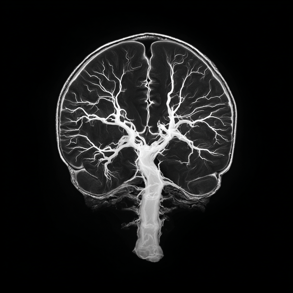
A brain MRI is a powerful window into the human mind, capable of revealing the intricate details of our neural landscape. For millions of people, a routine scan or one ordered for symptoms like headaches or dizziness may reveal a seemingly mysterious finding: “white matter changes.” In the radiology report, these may be called “white matter hyperintensities,” “leukoaraiosis,” or “small vessel ischemic disease.“ This diagnosis can be alarming and confusing, as it is often not explained in detail. However, these changes are a critical sign of a deeper process occurring in the brain and are strongly linked to an increased risk of stroke and a decline in memory and thinking abilities.
This article will decode the complex science behind white matter changes, explaining what they are, why they happen, their profound implications for both stroke and cognitive health, and the actionable steps you can take to manage and slow their progression.

What Is White Matter?
To understand white matter changes, it’s essential to first understand what white matter is. Imagine your brain as a massive, complex city. The gray matter is the city itself; the bustling hubs of activity where all the thinking, processing, and decision-making happen. The white matter is the vast network of roads, highways, and electrical wiring that connects all these hubs, allowing them to communicate with one another.
White matter is made up of millions of nerve fibers, or axons, that are insulated by a fatty substance called myelin. This myelin sheath acts like the rubber insulation on an electrical wire, allowing nerve signals to travel quickly and efficiently from one part of the brain to another. When this insulation or the underlying nerve fibers are damaged, the communication network begins to falter.
What Are White Matter Changes?
When a doctor refers to “white matter changes” on an MRI, they are looking at specific bright spots on the scan. These spots, known as hyperintensities, appear on a T2-weighted or FLAIR MRI sequence. On the scan, the healthy white matter appears dark, but areas that are damaged or have a buildup of fluid appear as bright, glowing spots. [2]
These changes are not a disease in themselves but rather a sign of an underlying process of damage. The presence of these bright spots indicates that the myelin and a small portion of the surrounding tissue have been injured. The number, size, and location of these hyperintensities are a measure of the severity of this damage.
Why Do White Matter Changes Happen?
The most common and significant cause of white matter changes is a condition called chronic cerebral ischemia: a persistent, low-grade lack of blood flow to the brain’s deep white matter. Think of it as a constant, low-level traffic jam in the brain’s circulatory system. This is typically a result of damage to the brain’s smallest arteries, a condition known as small vessel disease. [3]
The primary culprits behind this small vessel disease are the very same risk factors that lead to heart attacks and strokes:
- High Blood Pressure (Hypertension): Uncontrolled high blood pressure is the single most important risk factor for white matter changes. The constant force of the blood flow damages the tiny, delicate blood vessels in the brain, leading to a breakdown of the surrounding tissue. [4]
- Diabetes: High blood sugar levels can damage blood vessels throughout the body, including the small vessels in the brain.
- High Cholesterol: The buildup of fatty plaque in the arteries (atherosclerosis) can narrow or block the small blood vessels, restricting blood flow.
- Smoking: Smoking directly damages blood vessels and impairs the brain’s ability to get enough oxygen and nutrients.
- Aging: While not a disease, the natural aging process makes blood vessels more brittle and less resilient to damage. The presence of white matter changes is far more common in older adults. [5]
Other factors, such as migraines with aura and certain autoimmune diseases, have also been linked to white matter changes, but vascular risk factors remain the most important drivers.
A Warning Sign of Vascular Risk
For a patient and their doctor, the presence of white matter changes on an MRI should be considered a significant warning sign for future stroke risk. These changes are a tangible manifestation of small vessel disease, a process that weakens the brain’s tiny arteries and makes them highly susceptible to two types of stroke:
- Ischemic Stroke: The weakened vessels are prone to developing blockages from blood clots, cutting off blood supply to a portion of the brain and causing a stroke. This is particularly relevant for a type of ischemic stroke called a lacunar stroke, which occurs in the deep white matter where these changes are most prominent. [6]
- Hemorrhagic Stroke: The weakened vessels can also be more prone to rupturing, leading to a bleed in the brain.
Numerous studies have demonstrated a direct link between the volume and severity of white matter changes and the risk of stroke. The presence of these changes is a more reliable indicator of stroke risk than many other commonly used markers.
When the “Wiring” Fails
Beyond the risk of a catastrophic event like a stroke, white matter changes have a profound and progressive impact on cognitive function, particularly memory and thinking speed. While a person with a few small spots may not notice a difference, as the damage accumulates, the consequences become more apparent.
The damage to the brain’s white matter disrupts the efficient communication between different brain regions. This can lead to:
- Slowing of Thought: It takes longer for signals to travel between brain regions, leading to a general slowing of a person’s mental processing speed.
- Executive Dysfunction: This is the most common cognitive symptom. Executive function refers to the brain’s ability to plan, organize, make decisions, and manage tasks. White matter changes can make these complex processes more difficult. [7]
- Memory Impairment: While not the same as Alzheimer’s disease, WMC can affect memory, particularly the ability to retrieve information. They are a primary cause of vascular dementia, the second most common type of dementia after Alzheimer’s.
- Mixed Dementia: It is common for people to have both Alzheimer’s disease pathology and vascular brain changes (WMC). This is known as mixed dementia, and the vascular damage can worsen the cognitive symptoms of Alzheimer’s.
How to Manage White Matter Changes
The finding of white matter changes is not a cause for despair but a powerful call to action. While there is no pill or surgery that can reverse the damage, the key is to prevent it from getting worse. The primary treatment is aggressive management of the underlying vascular risk factors.
- Control Blood Pressure: This is the single most important step. Work with your doctor to maintain a healthy blood pressure, ideally below 120/80 mmHg. This is the most effective way to slow the progression of white matter changes. [9]
- Manage Diabetes and Cholesterol: Keep your blood sugar and cholesterol levels within a healthy range through diet, exercise, and medication as prescribed.
- Quit Smoking: Smoking cessation is a critical step to halting further damage to your blood vessels.
- Embrace a Healthy Lifestyle: Regular physical activity (aerobic exercise, like brisk walking), a heart-healthy diet (like the Mediterranean or DASH diet), and maintaining a healthy weight can all help improve blood flow and protect your brain. [10]
White Matter Changes in Perspective
White matter changes on an MRI are a diagnostic finding that points to a silent but progressive form of brain damage. They are not a natural part of aging but are a direct consequence of a lifetime of vascular stress, with hypertension being the main culprit. These changes are a powerful indicator of increased risk for both stroke and cognitive decline. However, they are also a wake-up call. You can effectively slow down the progression of this damage and protect your brain’s vital communication network, reducing your risk of future stroke and preserving your memory and cognitive function for years to come.
