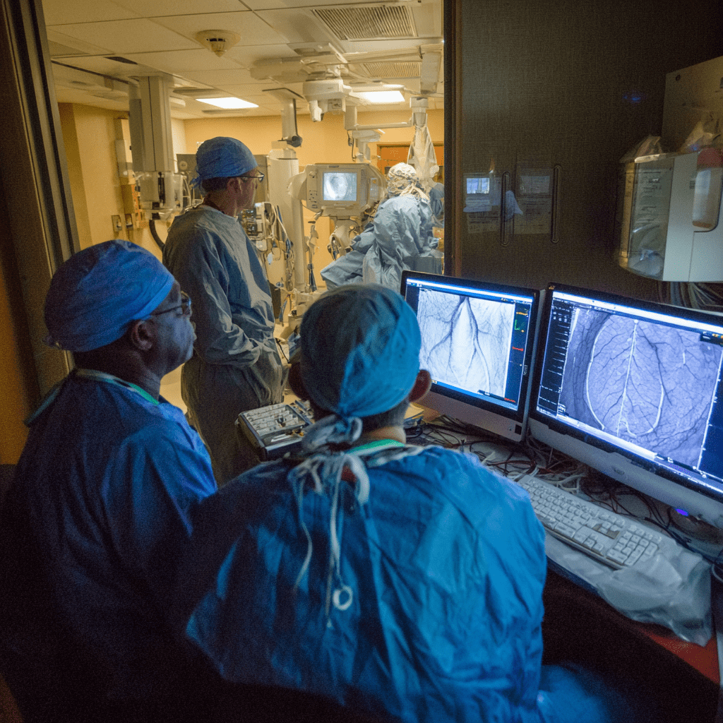
An angiogram is a sophisticated medical procedure that provides doctors with a detailed look inside the body’s blood vessels. By using a specialized dye and X-ray imaging, this test can reveal blockages, narrowings, aneurysms, and other abnormalities that can’t be seen with standard diagnostic tools. It is considered the gold standard for diagnosing a range of cardiovascular conditions, most notably coronary artery disease. While the prospect of a medical procedure can be intimidating, understanding each step, from preparation to recovery, can alleviate anxiety and empower patients. This article will talk you through the angiogram procedure, detailing its purpose, the step-by-step process, potential risks, and what to expect during recovery.

What Is an Angiogram and Why Is It Performed?
An angiogram is an X-ray examination of blood vessels, including arteries and veins. The term itself is derived from the Greek words angeion (vessel) and graphein (to write or record). The procedure is performed in a hospital’s catheterization lab by a cardiologist or a radiologist.
The primary purpose of an angiogram is to diagnose the location and severity of blood vessel issues. A doctor may recommend an angiogram to:
- Identify Blockages: Pinpoint the exact location of fatty plaque buildup (atherosclerosis) in the arteries, which can lead to a heart attack or stroke.
- Assess Blood Flow: Evaluate how well blood is flowing to specific organs, such as the heart, brain, or legs.
- Diagnose Aneurysms: Identify a weakened or bulging section of a blood vessel that could rupture.
- Determine Treatment: The results of an angiogram are often used to guide a subsequent procedure, such as angioplasty (to open a blocked artery) or the placement of a stent. It is also essential for planning surgical interventions like coronary artery bypass grafting. [1]
While angiograms can be performed on blood vessels throughout the body, the most common is the coronary angiogram, which visualizes the arteries supplying blood to the heart. Other types include cerebral angiography (for the brain) and peripheral angiography (for the legs).
The Angiogram Procedure
The angiogram is a minimally invasive procedure, typically performed with the patient awake but sedated. While the experience can vary slightly depending on the specific blood vessels being examined, the general process follows a consistent sequence.
1. Patient Preparation
Prior to the procedure, you will be given specific instructions. This usually involves:
- Fasting for several hours before the test to prevent nausea.
- Stopping certain medications, particularly blood thinners, as advised by your doctor. [2]
- An IV line will be placed in your arm to administer fluids and sedation.
2. The Catheterization Lab
You will be moved to a sterile environment called a catheterization lab. You’ll lie on a special table, and a team of doctors, nurses, and technicians will be present. Your heart rate, blood pressure, and oxygen levels will be monitored throughout the procedure.
3. Access Site Preparation
The doctor will choose an access site to insert the catheter. The most common sites are the femoral artery in the groin or the radial artery in the wrist. The chosen area will be shaved, cleaned with an antiseptic solution, and then injected with a local anesthetic to numb the skin and underlying tissue. [3]
4. Catheter Insertion
Once the area is numb, a small incision, typically less than a quarter of an inch, is made. A short, hollow sheath is inserted into the artery. A long, thin, flexible tube called a catheter is then threaded through this sheath. Using a live X-ray screen (fluoroscopy) as a guide, the doctor carefully navigates the catheter through the blood vessels to the targeted area. You will not feel the catheter moving through your vessels.
5. Dye Injection and Imaging
When the catheter is in the correct position, a contrast dye is injected. This dye, which is visible on the X-ray, makes the blood vessels appear as dark outlines on the monitor. You may feel a warm, flushing sensation as the dye travels through your body, but this is a normal and temporary sensation. As the dye flows, the doctor takes a series of X-ray images or a video recording to observe the blood flow and identify any narrowings or blockages. The contrast dye is what allows the doctor to see precisely where a problem is located. [5]
6. Catheter Removal and Closure
Once the necessary images have been obtained, the catheter and sheath are carefully removed. The doctor will apply firm pressure to the incision site for several minutes to stop any bleeding. A dressing will then be applied. In some cases, a special closure device may be used to seal the artery and minimize bleeding. [6]
Understanding the Risks and Complications
An angiogram is considered a safe procedure, and the benefits of an accurate diagnosis almost always outweigh the risks. However, as with any medical procedure, there are potential complications.
Common Risks:
- Bleeding or Bruising: The most frequent complication is bleeding or bruising at the catheter insertion site. This is usually minor and resolves on its own within a few days. [7]
- Discomfort or Pain: You may feel some soreness at the incision site.
Rare but Serious Risks:
- Allergic Reaction to the Dye: Some people may have an allergic reaction to the contrast dye, ranging from a mild rash to a severe reaction that affects breathing.
- Kidney Damage: The contrast dye can sometimes cause temporary or permanent damage to the kidneys, particularly in patients with pre-existing kidney disease or diabetes. To mitigate this risk, patients are encouraged to stay well-hydrated before and after the procedure to help flush the dye from their system. [8]
- Artery Damage: In very rare instances, the catheter can damage the artery or cause a blood clot.
- Stroke or Heart Attack: While extremely rare, there is a minute risk that the procedure could dislodge a piece of plaque, leading to a stroke or heart attack.
- Infection: Any procedure that involves breaking the skin carries a small risk of infection.
The Recovery Journey
Your recovery is just as important as the procedure itself to ensure a smooth outcome.
Immediately After the Procedure:
You will be taken to a recovery area for close monitoring. The most critical part of this stage is to prevent bleeding from the access site.
- Groin Access: If the femoral artery in your groin was used, you will be required to lie flat on your back for several hours, with minimal movement, to allow the artery to seal. [9]
- Wrist Access: If the radial artery in your wrist was used, a special compression band will be applied. This allows for earlier mobilization.
At Home:
- Rest: Plan to rest for at least 24 hours after the procedure. Avoid any strenuous activity, heavy lifting, or intense exercise for a few days, as advised by your doctor.
- Hydration: Drink plenty of water to help your kidneys flush out the remaining contrast dye.
- Monitor the Site: Keep the bandage on as instructed. Watch for signs of complications, such as swelling, spreading redness, severe pain, or bleeding. If a large, painful lump develops, or the site starts to bleed heavily, seek immediate medical attention. [10]
Your doctor will discuss the results of the angiogram with you and your family, explaining what was found and outlining the next steps, whether that’s medication, lifestyle changes, or a follow-up procedure.
