Introduction
The scenario is a familiar one: a seemingly routine ankle sprain that just won’t heal. You’ve rested, iced, and elevated your foot, but weeks or even months later, a nagging, deep-seated pain and a feeling of instability persist. While the initial X-ray was likely normal, your doctor may suggest an MRI. This is often the critical turning point in diagnosing a condition that a simple sprain cannot explain: an osteochondral lesion (OCL) of the ankle.
An OCL is a type of injury that affects both the cartilage and the underlying bone of a joint. It is a common cause of chronic ankle pain, but it is frequently missed in initial examinations. This article will demystify what an ankle osteochondral lesion is, explain why it often mimics a sprain, and, most importantly, highlight the crucial role of an MRI in revealing the true extent of the damage and guiding the path to effective treatment.

What is an Ankle Osteochondral Lesion?
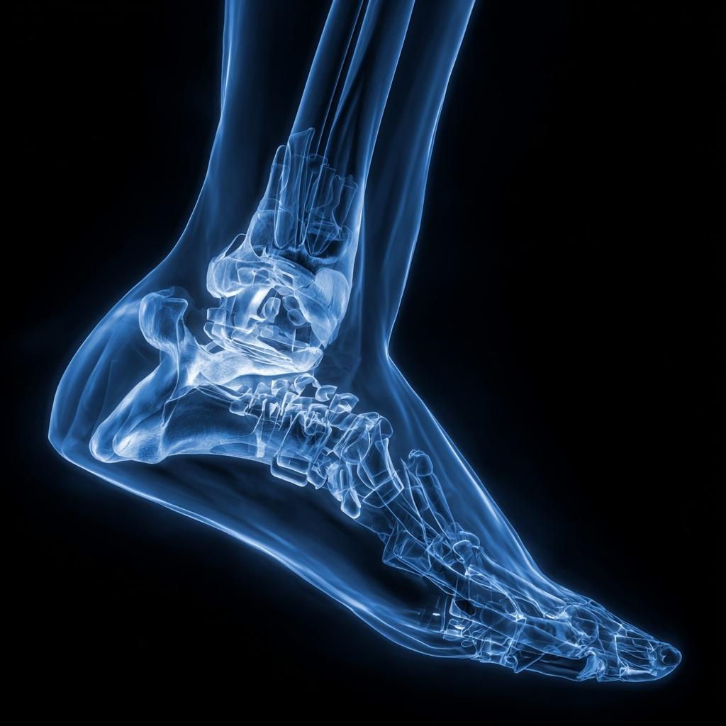
To understand an OCL, you must first understand the anatomy of the ankle joint. The ankle is a hinge joint formed by three bones: the tibia (shin bone), the fibula (the smaller leg bone), and the talus (the ankle bone). The top of the talus is shaped like a dome and is covered by a smooth layer of articular cartilage. This cartilage acts as a shock absorber and allows the bones to glide effortlessly against each other. [1]
An osteochondral lesion (OCL) is a localized injury to this articular cartilage and the underlying subchondral bone. The injury can range in severity from a simple bruise to a full-thickness defect where a piece of the bone and cartilage has completely broken off and is floating freely in the joint. The most common cause of an OCL is an acute traumatic event, such as a severe ankle sprain or a fracture. During a forceful twisting of the ankle, the talus bone can violently impact the tibia, causing the cartilage and bone to compress or shear off. [2] They can also be caused by chronic, repetitive microtrauma or a condition called avascular necrosis, where the bone loses its blood supply and dies.
Why They Mimic a Sprain
The symptoms of an OCL are often vague and can be easily confused with a persistent ankle sprain, which is why they are so commonly misdiagnosed.
- Persistent, Deep Pain: The pain is often described as a deep ache within the ankle joint. It is typically a dull, persistent pain that worsens with weight-bearing activities like walking, running, or standing for long periods.
- Chronic Swelling: Persistent swelling that doesn’t resolve after the typical healing period for a sprain is a key indicator.
- Instability and “Giving Way”: The joint may feel unstable or feel like it is “giving way,” particularly on uneven ground.
- Locking, Catching, or Clicking: If a piece of bone or cartilage has broken off and is floating in the joint, it can cause a “locking” or “catching” sensation when you move your ankle. [3]
The Diagnostic Power of MRI
When a patient’s pain lingers despite conservative treatments for a sprain, a doctor will often order an MRI. The MRI is the definitive diagnostic tool for an OCL because it can visualize both soft tissue (cartilage) and bone, providing a level of detail that a standard X-ray cannot match. A simple X-ray can only show bone and will almost always miss a cartilage injury.
Here is what a radiologist and an orthopedic surgeon are looking for on an MRI report:
- Bone Bruise (Edema): The MRI can show swelling and fluid within the talus bone, indicating that a significant impact has occurred. This is often the first sign of an OCL. [4]
- Cartilage Integrity: The MRI provides a clear picture of the articular cartilage. It can identify if the cartilage is thin, cracked, or if there is a full-thickness defect where the cartilage is completely gone. This helps determine the stage and severity of the lesion.
- Loose Fragments: The MRI can clearly show if a piece of bone and cartilage has broken off and is now a “loose body” floating in the joint space. This is a critical finding that often necessitates surgical intervention. [5]
- Bone Cyst Formation: The MRI can also reveal if a subchondral bone cyst has formed beneath the lesion, which indicates a more chronic injury and helps guide the treatment plan.
Treatment Based on MRI Findings
The detailed information provided by the MRI is what allows a doctor to formulate an accurate and effective treatment plan.
- Conservative Treatment: For small, stable OCLs where the MRI shows a bone bruise but the cartilage is intact, the first line of treatment is typically conservative. This includes a period of rest, immobilization in a boot, and physical therapy. The goal is to allow the bone and cartilage to heal on their own.
- Surgical Intervention: For larger, unstable, or symptomatic OCLs, surgery is often necessary. The MRI helps the surgeon plan the specific procedure, which can range from a minimally invasive arthroscopic procedure to a more complex open surgery. Surgical options may include:
- Debridement: Removing any loose fragments and smoothing the rough edges of the lesion.
- Microfracture: A procedure to stimulate new cartilage growth.
- Osteochondral Autograft Transplantation (OAT): A more advanced procedure that involves transplanting healthy cartilage and bone from another part of the body to the injured site. [6]
