Introduction
Receiving an MRI report that mentions “brain bleeds” or “microhemorrhages” can be a deeply alarming experience. The term itself conjures images of a major, life-threatening stroke. However, in most cases, microhemorrhages are not a sign of an active, dangerous bleed. Instead, they are tiny, chronic remnants of past bleeding, often appearing as nothing more than minute black dots on a specific type of MRI scan. Their significance is not in their size but in their number, location, and the underlying condition that caused them. We will provide a clear and comprehensive overview of microhemorrhages, helping you understand their causes, their clinical significance, and what they might mean for your long-term health.

What Are Microhemorrhages?
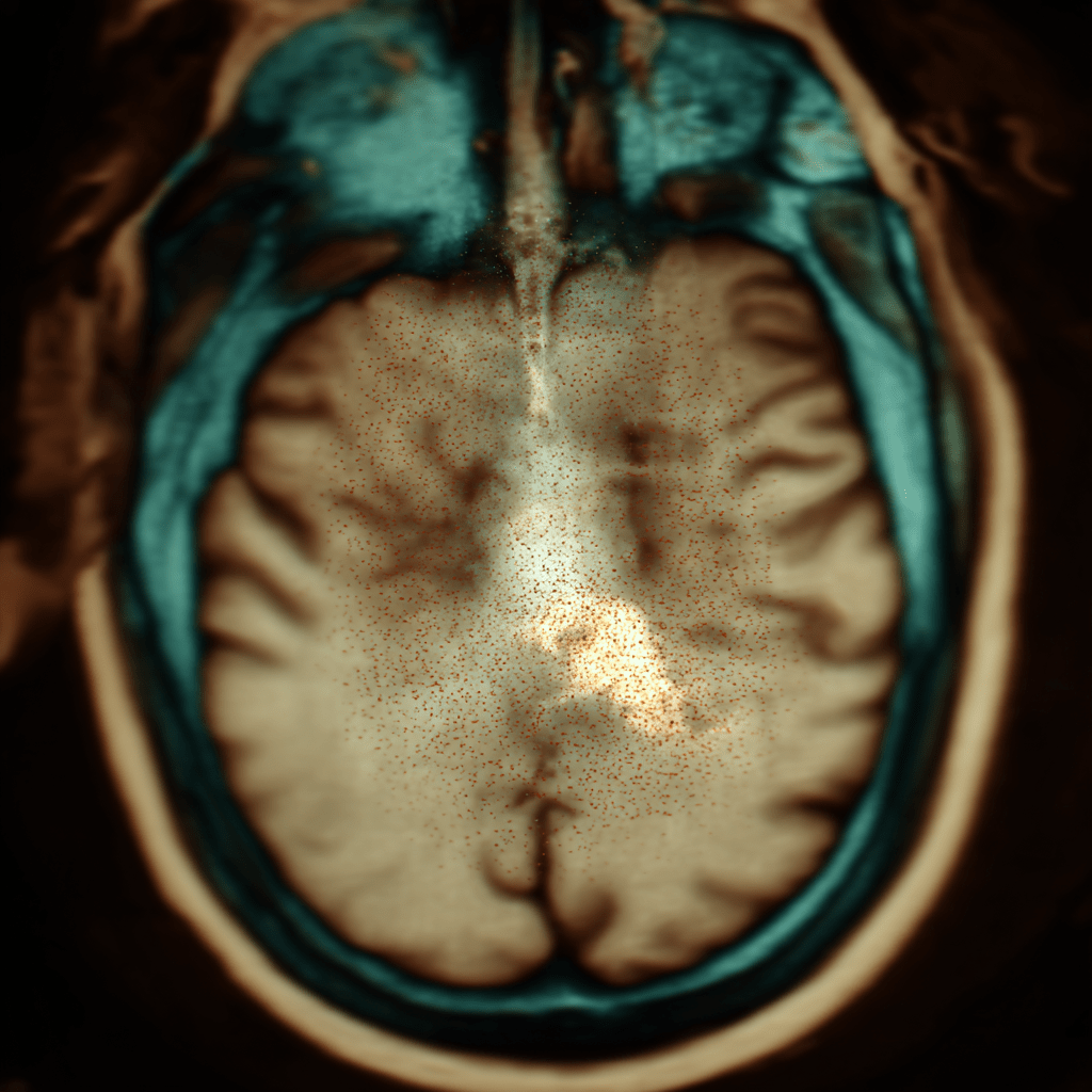
Microhemorrhages, also known as cerebral microbleeds (CMBs), are small, chronic deposits of blood breakdown products in the brain. They are the leftover traces of old, microscopic hemorrhages from tiny, fragile blood vessels. These bleeds are too small to cause symptoms like a major stroke and are invisible on a standard CT scan or a regular MRI sequence. Instead, they are detected using a highly sensitive MRI technique called susceptibility-weighted imaging (SWI) or a similar sequence. On these scans, the iron within the blood deposits creates a magnetic artifact that makes them appear as distinct, tiny black dots, typically a few millimeters in size. [1] Their presence is a telltale sign that there is an underlying issue with the brain’s small blood vessels.
The Main Causes of Microhemorrhages
Microhemorrhages are not a disease in themselves but a sign of a broader problem with the brain’s vascular system. Their cause is often linked to their location in the brain. The two most common causes are hypertension and cerebral amyloid angiopathy (CAA).
1. Hypertensive Microangiopathy
Chronic, uncontrolled high blood pressure is the leading cause of deep-seated microhemorrhages. Over time, high blood pressure damages the very small arteries that penetrate deep into the brain. This damage makes the vessel walls weak and leaky, leading to tiny, slow hemorrhages. These microbleeds are typically found in the subcortical regions of the brain, such as the basal ganglia, thalamus, and brainstem. [2]
2. Cerebral Amyloid Angiopathy (CAA)
This is a condition where a protein called amyloid-beta builds up in the walls of the blood vessels in the brain. This amyloid accumulation makes the vessels brittle and fragile, significantly increasing the risk of both microscopic and larger, symptomatic hemorrhages. Microhemorrhages related to CAA are typically found in the outer layers of the brain, known as the cortex or lobar regions, and are strongly associated with Alzheimer’s disease. [3]
3. Other Causes
While less common, other factors can also lead to microhemorrhages:
- Traumatic Brain Injury (TBI): Especially in severe cases, a TBI can cause widespread damage to blood vessels, leading to microbleeds.
- Medications: The use of blood-thinning medications, especially in older adults, can increase the risk of both micro- and macro-hemorrhages. [4]
- Genetic Disorders: Certain rare genetic conditions can predispose individuals to fragile blood vessels and brain bleeds.
When Are They Harmless vs. Serious?
The mere presence of a microhemorrhage is not an immediate cause for alarm. Its significance is determined by a neurologist or radiologist who considers its location, number, and the patient’s overall clinical picture.
When They May Be Benign (Harmless)
- Small Number: A single or a very small number (e.g., one or two) of microhemorrhages, especially in an older person with no other symptoms or risk factors, may be considered an age-related finding and have little immediate clinical significance. [5]
- Location: Deep-seated bleeds are a sign of small vessel disease related to hypertension, but they are not as strongly linked to large, catastrophic bleeds as CAA-related hemorrhages. If a patient’s blood pressure is well-controlled, the risk may be minimal.
When They Are a Serious Red Flag
- High Number: A large number of microhemorrhages (e.g., more than 5 to 10) is a significant concern and a clear sign of widespread small vessel disease. This indicates a higher risk for future strokes and cognitive impairment.
- Location: Microbleeds in the lobar (cortical) regions of the brain are highly suggestive of Cerebral Amyloid Angiopathy. This finding is particularly worrisome as it is a strong predictor of a future lobar hemorrhage (a type of hemorrhagic stroke) and is often an early sign of Alzheimer’s-related brain changes. [7]
- Associated Symptoms: The most important factor. If microhemorrhages are found in a patient with a history of stroke, transient ischemic attack (TIA), cognitive impairment, or memory problems, they are no longer a benign finding. In this context, they are likely contributing to the person’s symptoms and are a sign of progressive neurological disease.
What to Do About Microhemorrhages
Receiving a diagnosis of microhemorrhages is not a death sentence; it is an important piece of information that can inform your treatment and lifestyle to protect your future health.
- Identify the Cause: The first step is for your doctor to determine the underlying cause. An MRI scan can help differentiate between hypertension-related and CAA-related bleeds based on their location, but additional tests may be needed.
- Aggressive Blood Pressure Management: If hypertension is the cause, the most effective strategy is to aggressively manage your blood pressure. Consistent blood pressure control is the single best way to prevent further damage to your blood vessels and reduce your risk of future strokes. [8]
- Medication Review: If you are on blood-thinning medication (e.g., anticoagulants or antiplatelets), your doctor will need to carefully weigh the risks and benefits of continued use. In some cases, the risk of a new brain bleed may outweigh the benefit of preventing a blood clot. [9]
- Lifestyle Modifications: Regardless of the cause, a heart-healthy lifestyle is a brain-healthy lifestyle. This includes a balanced diet, regular exercise, and not smoking.
- Cognitive Monitoring: For patients with a high burden of microhemorrhages, especially those in the cortex, regular cognitive assessments may be recommended to monitor for any signs of cognitive decline.
