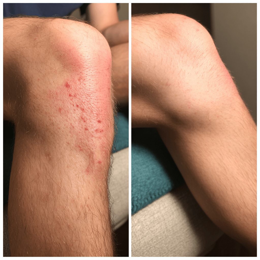
The knee joint, a complex articulation essential for mobility, is susceptible to various pathologies, one of the most common being a joint effusion. Specifically, a suprapatellar joint effusion refers to the accumulation of excess fluid within the suprapatellar bursa, a synovial sac located superior to the patella. This condition, colloquially known as “water on the knee,” is a clinical sign of an underlying issue rather than a primary diagnosis. Understanding its anatomical context, etiology, and the evidence-based approaches to its management is crucial for effective treatment and patient recovery.

Anatomical and Physiological Basis
The knee is a synovial joint, meaning it is encased in a capsule that contains synovial fluid. This viscous fluid serves a critical function, providing lubrication to the articular cartilage, reducing friction during movement, and supplying nutrients to the avascular cartilage. The synovial capsule is not a simple, single compartment; it includes several recesses and extensions. The suprapatellar bursa (or pouch) is a crucial extension located anteriorly, between the distal quadriceps tendon and the anterior surface of the distal femur.
In most individuals, the suprapatellar bursa communicates freely with the main knee joint cavity, allowing fluid to move between these spaces. [2] This communication is vital for distributing synovial fluid during knee flexion and extension. An effusion, therefore, is not limited to the suprapatellar bursa but represents an increase in the total volume of intra-articular fluid. This fluid is a plasma ultrafiltrate, containing hyaluronic acid, glycoproteins, and other substances that maintain joint health. Under pathological conditions, the synovium becomes inflamed, leading to increased vascular permeability and the overproduction of synovial fluid, a process that can cause significant distension of the suprapatellar bursa.
Etiology and Pathophysiology
The causes of a suprapatellar joint effusion are multifactorial and can be broadly categorized into traumatic, inflammatory, degenerative, and infectious conditions.
Traumatic Causes:
Acute trauma is a frequent cause, often leading to a rapid accumulation of fluid, which may contain blood (hemarthrosis). Common traumatic injuries include:
- Ligamentous Tears: A tear of the anterior cruciate ligament (ACL) is a primary cause of hemarthrosis due to the rich vascularity of the ligament. [4]
- Meniscal Tears: The menisci, C-shaped fibrocartilaginous pads, can tear from twisting injuries, leading to joint irritation and a subsequent effusion.
- Fractures: Intra-articular fractures, such as those of the tibial plateau or femoral condyles, can cause both an effusion and hemarthrosis due to bone marrow hemorrhage into the joint space.
Inflammatory and Degenerative Causes:
These conditions typically result in a more gradual onset of swelling. The effusion in these cases is primarily inflammatory fluid, rich in white blood cells and protein.
- Osteoarthritis (OA): A degenerative joint disease, OA is characterized by the breakdown of articular cartilage. The body’s inflammatory response to the cartilaginous debris and joint space narrowing leads to chronic synovial inflammation and an effusion.
- Rheumatoid Arthritis (RA): This is a systemic autoimmune disease where the body’s immune system mistakenly attacks the synovial membrane, causing chronic inflammation, pain, and a persistent effusion.
- Crystal Arthropathies: Conditions like gout and pseudogout are caused by the deposition of crystals (uric acid in gout, calcium pyrophosphate dihydrate in pseudogout) in the joint, triggering an intense inflammatory reaction and a painful effusion.
Infectious Causes:
Septic arthritis, or a joint infection, is a medical emergency. Bacteria, viruses, or fungi can enter the joint space, leading to a severe inflammatory response and rapid fluid accumulation. The effusion fluid, in this case, is purulent (pus-like) and requires immediate medical attention to prevent irreversible joint damage.
Clinical Presentation and Diagnosis
The clinical presentation of a suprapatellar joint effusion includes visible swelling of the knee, a loss of the normal knee contours, and a palpable fluid bulge above the kneecap. Patients often report a sensation of tightness or pressure and a reduced range of motion. The presence of pain and warmth further suggests an inflammatory or infectious process.
Diagnosis relies on a combination of a thorough clinical examination and diagnostic imaging.
Physical Examination:
A clinician will perform specific tests to confirm the presence of an effusion. The Patellar Tap Test (or “ballottement”) involves applying downward pressure on the suprapatellar pouch and then tapping the patella. If an effusion is present, the patella will be felt to “bounce” or “tap” against the femur. The Bulge Sign (or “sweep test”) is used for smaller effusions and involves milking fluid from one side of the joint to the other, creating a palpable or visible bulge. [9]
Imaging Studies:
- X-rays: are primarily used to assess for bone abnormalities such as fractures, osteophytes (bone spurs), or signs of arthritis. An effusion may be visible as a soft tissue density and displacement of the suprapatellar fat pad on a lateral view.
- Ultrasound: is an excellent tool for visualizing and quantifying an effusion in real-time. It can identify the presence of fluid, guide aspiration procedures, and differentiate between a simple effusion and a more complex fluid collection.
- Magnetic Resonance Imaging (MRI): provides a detailed view of the soft tissues, including the ligaments, menisci, and cartilage. It is the gold standard for identifying the underlying cause of an effusion, such as a ligament tear or meniscal injury.
Arthrocentesis (Joint Aspiration):
This procedure involves inserting a needle into the joint space to withdraw fluid. The aspirated fluid can be analyzed for various parameters, including cell count, protein levels, and the presence of crystals or bacteria. This is particularly critical in cases where septic arthritis is suspected.
Treatment and Management Strategies
The management of a suprapatellar joint effusion is centered on addressing the underlying cause while also alleviating symptoms. A tiered approach, progressing from conservative to more invasive methods, is typically employed.
1. Conservative Management:
- RICE Protocol: This is a cornerstone of initial management for traumatic causes. Rest helps prevent further injury, Ice application reduces inflammation and pain, Compression with an elastic bandage can help control swelling, and Elevation of the limb above the heart facilitates fluid drainage via gravity. [13]
- Medications: Non-steroidal anti-inflammatory drugs (NSAIDs) such as ibuprofen or naproxen are effective for reducing pain and inflammation associated with non-infectious effusions.
- Activity Modification: Avoiding high-impact activities and movements that exacerbate symptoms is crucial during the acute phase of recovery.
2. Physical Therapy:
Once the acute swelling has subsided, physical therapy is essential for restoring joint function and preventing muscle atrophy. A comprehensive program may include:
- Range of Motion Exercises: Gentle flexion and extension exercises help maintain joint mobility.
- Strengthening Exercises: Strengthening the quadriceps, hamstrings, and gluteal muscles provides better support and stability for the knee joint, which can reduce the risk of future effusions.
- Balance and Proprioception Training: These exercises help retrain the body’s sense of joint position, which is often impaired after a knee injury or swelling. [14]
3. Interventional Procedures:
- Arthrocentesis: While a diagnostic tool, it is also a therapeutic procedure. Aspiration of a large, tense effusion can provide immediate relief from pain and pressure.
- Corticosteroid Injections: Following aspiration, a corticosteroid may be injected into the joint to reduce local inflammation. This is particularly effective for inflammatory conditions like osteoarthritis or RA and can provide long-lasting relief, though it is not a cure. [15] These injections are not recommended for infectious effusions.
4. Surgical Intervention:
Surgical treatment is reserved for cases where conservative and interventional methods are insufficient or when the underlying cause requires repair.
- Arthroscopy: A minimally invasive procedure where a surgeon uses a small camera and instruments to visualize and treat the joint. Arthroscopy can be used to repair a torn meniscus, reconstruct a torn ligament, or debride inflamed synovial tissue (synovectomy). [16]
- Total Knee Arthroplasty (TKA): In severe cases of end-stage arthritis where the joint is completely degenerated, a knee replacement may be the definitive treatment to eliminate the source of the chronic effusion and restore function.
