
You go for a MRI scan because of occasional back discomfort, stiffness, or perhaps a mild ache that has been bothering you for a few weeks. You are still walking comfortably, managing your daily activities, and sleeping reasonably well. Nothing about your symptoms seems particularly serious.
Then the MRI report arrives.
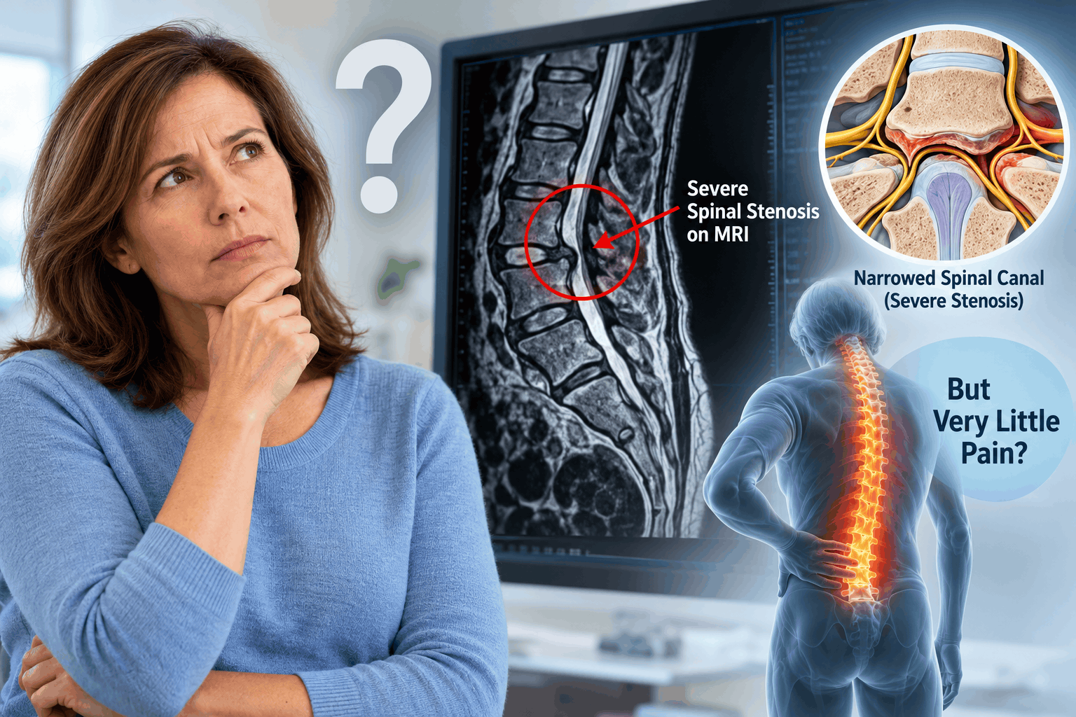
It mentions severe spinal canal stenosis, significant narrowing around the nerves, or advanced degenerative changes in the spine.
Naturally, the word “severe” raises concerns. How can something described as severe on an MRI cause so little pain? Does it mean your spine is deteriorating without you realizing it? Could you suddenly develop nerve damage? And perhaps most importantly, do you need surgery even though you feel relatively fine?
The surprising answer is that the severity of spinal stenosis on an MRI does not always correspond to the amount of pain or disability a person experiences.
Some individuals have considerable narrowing of the spinal canal but remain physically active with minimal symptoms. Others experience substantial leg pain, numbness, or difficulty walking despite having only moderate narrowing on imaging.
This difference is well recognized in medical research. A 2023 publication discussing clinical practice guidelines for lumbar spinal stenosis emphasized that the severity of narrowing seen on magnetic resonance imaging does not consistently correlate with pain intensity, walking distance, or quality of life.
Understanding why this happens can help patients interpret their MRI reports more accurately and avoid unnecessary anxiety or treatment.
What Does Severe Spinal Stenosis Actually Mean on an MRI?
Spinal stenosis refers to narrowing of the spaces within the spine through which the spinal cord or spinal nerves travel.
The spinal column consists of vertebrae, intervertebral discs, joints, ligaments, and other supporting structures. Together, these structures protect the nervous system while allowing the back and neck to move.
Over time, several age-related changes can reduce the available space around the nerves.
These changes include:
- Bulging or degenerating intervertebral discs.
- Enlargement of the facet joints.
- Thickening of the ligamentum flavum.
- Formation of bone spurs.
- Degenerative spondylolisthesis, where one vertebra slips relative to another.
- Arthritic changes involving multiple spinal structures.
When these changes significantly reduce the available space, the radiologist may describe the condition as severe spinal stenosis.
However, there is an important distinction.
Severe spinal stenosis describes the degree of anatomical narrowing. It does not automatically describe the severity of a patient’s symptoms.
A radiologist evaluates what the images show. A treating physician must determine whether those findings are responsible for the patient’s symptoms, whether neurological function is affected, and whether treatment is necessary.
What Is the Difference Between Mild, Moderate, and Severe Spinal Stenosis?
Radiologists generally classify spinal stenosis according to the amount of narrowing and the appearance of the structures surrounding the nerves.
In mild spinal stenosis, there is some narrowing, but adequate space generally remains around the neural structures.
Moderate spinal stenosis involves more noticeable narrowing, sometimes accompanied by crowding of the nerve roots.
Severe spinal stenosis indicates substantial narrowing, potentially with marked crowding or compression of neural structures.
The precise grading criteria vary depending on whether the narrowing affects the central spinal canal, lateral recess, or neural foramen.
A severe finding at one spinal level may also have a different clinical significance from narrowing at another level.
For example, severe lumbar spinal stenosis primarily affects nerve roots, while severe cervical spinal stenosis may compress the spinal cord itself.
This distinction becomes particularly important when deciding whether a patient requires further evaluation or treatment.
Why Can Severe Spinal Stenosis Cause Very Little Pain?
There is no single explanation for why someone with severe spinal stenosis may experience minimal discomfort.
Pain is influenced by several factors, including nerve irritation, inflammation, mechanical loading, individual nervous system responses, and the location of the narrowing.
1. Spinal Canal Narrowing Does Not Always Produce Significant Nerve Irritation
One of the most important reasons for the mismatch between MRI findings and symptoms is that anatomical narrowing does not necessarily produce substantial nerve irritation.
A nerve may have less surrounding space without being irritated enough to generate significant pain.
Consider two individuals with similarly severe lumbar spinal stenosis.
One experiences burning pain extending into the legs after walking for several minutes.
The other notices only mild stiffness after prolonged standing.
Although their MRI reports may appear similar, the functional effects on their nerves can be different.
Factors such as inflammation, local pressure, blood flow, and the particular nerve structures involved may influence symptoms.
This helps explain why a measurement of spinal canal narrowing cannot reliably predict an individual’s pain level.
A 2020 review by the World Federation of Neurosurgical Societies Spine Committee concluded that the degree of stenosis seen on imaging does not reliably correspond to the severity of clinical complaints.
2. Spinal Stenosis Usually Develops Gradually
Degenerative spinal stenosis commonly develops over several years.
Intervertebral discs gradually lose height, facet joints enlarge, and ligaments may become thicker.
Because these changes often occur slowly, symptoms may develop gradually rather than appearing suddenly.
Some people remain comfortable despite substantial anatomical narrowing.
However, gradual progression should not be interpreted as proof that the nerves have adapted safely or that future neurological problems cannot develop.
The important point is that anatomical changes and clinical symptoms do not necessarily progress at the same rate.
3. Pain Depends on More Than the Amount of Nerve Compression
Pain is not simply a direct measurement of how tightly a nerve is compressed.
The nervous system responds to mechanical pressure, chemical irritation, inflammation, and other physiological influences.
Two people with comparable anatomical narrowing may therefore experience very different symptoms.
For example, one person may have narrowing accompanied by nerve root inflammation, producing radiating pain.
Another may have similar narrowing without substantial inflammation or painful nerve irritation.
Differences in individual pain processing may also contribute to variations in symptoms.
This does not mean that pain is imaginary or that severe stenosis is harmless. It means that pain is a complex biological experience that cannot be predicted from spinal anatomy alone.
4. Your Daily Activities May Not Trigger the Stenosis
Many people with lumbar spinal stenosis experience symptoms primarily during specific activities.
Standing upright for prolonged periods or walking may increase discomfort, while sitting or bending forward may provide relief.
This happens partly because the dimensions of the lumbar spinal canal change with spinal position.
Lumbar extension, such as arching the lower back, can reduce the space available around the nerves.
Flexion, or bending forward, generally increases that space.
Someone who spends much of the day sitting, working at a desk, or performing activities that involve slight forward bending may experience fewer symptoms than someone who walks long distances or stands for several hours.
This can create the impression that severe spinal stenosis is completely asymptomatic.
However, symptoms may become more noticeable when the individual performs activities that place greater demands on the affected spinal region.
The relationship between posture, walking, and symptoms is a recognized feature of lumbar spinal stenosis.
5. Severe Narrowing May Affect an Area That Is Not Producing Symptoms
The location of spinal stenosis matters considerably.
Spinal narrowing can occur in several different regions.
Central canal stenosis involves narrowing of the main spinal canal.
Lateral recess stenosis affects the region through which nerve roots travel before exiting the spinal canal.
Foraminal stenosis involves narrowing of the openings through which spinal nerves leave the spine.
These different patterns can produce different symptoms.
For example, severe narrowing of a neural foramen may cause little discomfort if the affected nerve root is not significantly irritated.
Conversely, moderate narrowing in a location that places substantial pressure on a sensitive nerve root may produce considerable pain.
The presence of severe stenosis at multiple spinal levels also does not necessarily mean that every level is contributing to symptoms.
This is one reason physicians carefully compare MRI findings with the patient’s physical examination and distribution of symptoms.
Can You Have Severe Lumbar Spinal Stenosis Without Back Pain?
Yes. Severe lumbar spinal stenosis can occur with little or no back pain.
In fact, back pain is not always the dominant symptom of lumbar spinal stenosis.
Some patients primarily experience symptoms involving the buttocks or legs.
These may include:
- Leg heaviness during walking.
- Numbness or tingling in the feet.
- Aching in the buttocks or thighs.
- Weakness after prolonged standing.
- Reduced walking endurance.
- Symptoms that improve after sitting or leaning forward.
This pattern is often associated with neurogenic claudication, a characteristic manifestation of symptomatic lumbar spinal stenosis.
A person may report almost no lower back pain yet develop considerable difficulty walking.
For this reason, asking only about pain intensity can overlook important functional limitations.
A physician may also ask how far the patient can walk, whether they need to sit frequently, and whether their legs become weak or heavy during activity.
Why Walking Distance Can Matter More Than Back Pain
Consider a person who reports back pain of only 2 out of 10.
At first glance, the symptoms appear mild.
However, suppose the same individual can walk only 100 meters before experiencing significant leg heaviness and needing to sit.
Despite the relatively low pain score, spinal stenosis may be causing meaningful functional impairment.
Another person with an identical MRI report may walk several kilometers without difficulty.
These differences demonstrate why clinical evaluation must consider function rather than pain intensity alone.
A 2021 clinical review emphasized that treatment decisions for lumbar spinal stenosis should consider walking limitations, neurological deficits, and the overall impact on daily life.
Why Do Some People Have Severe Spinal Stenosis Without Any Symptoms?
Asymptomatic degenerative spinal changes are common, particularly with increasing age.
A well-known systematic review published in the American Journal of Neuroradiology examined imaging findings in 3,110 individuals who did not have symptoms.
The researchers found that degenerative spinal changes were frequently present even in people who were not experiencing pain.
For example, the estimated prevalence of disc degeneration increased from approximately 37% among asymptomatic 20-year-olds to 96% among asymptomatic 80-year-olds.
These figures concern disc degeneration rather than severe spinal stenosis specifically, but they illustrate an important principle: abnormalities seen on spinal imaging do not automatically indicate a painful medical condition.
The researchers concluded that many degenerative imaging findings are associated with normal aging and may not be responsible for symptoms.
Does This Mean Severe Spinal Stenosis Is a Normal Part of Aging?
Not exactly.
Age-related degeneration is common, but severe spinal stenosis should not automatically be dismissed as a harmless consequence of getting older.
The degree of narrowing, its location, the presence of spinal cord compression, and the patient’s neurological findings all matter.
Some individuals with severe stenosis remain stable for years.
Others develop increasing walking limitations, numbness, weakness, or neurological impairment.
Therefore, an asymptomatic MRI finding may not require immediate treatment, but it still deserves appropriate clinical interpretation.
Can an MRI Make Spinal Stenosis Look Worse Than It Really Is?
An MRI is highly useful for identifying anatomical narrowing, but it has limitations.
Most conventional spinal MRI examinations are performed while the patient is lying down.
This means the images generally represent the spine in a relatively unloaded position.
They do not necessarily show what happens during standing, walking, bending, or other activities.
In some patients, narrowing may become more pronounced during weight-bearing or spinal extension.
In others, the MRI may show substantial narrowing even though the affected nerves are not producing meaningful symptoms during everyday activities.
MRI Reports Also Involve Clinical Interpretation
Radiologists use established imaging characteristics to describe stenosis, but grading is not perfectly uniform.
Different specialists may occasionally classify the same degree of narrowing somewhat differently.
The 2023 lumbar spinal stenosis diagnostic guidelines discuss variability in imaging interpretation and emphasize that spinal stenosis should not be diagnosed solely from MRI findings without considering symptoms and physical examination.
This does not mean the MRI report is incorrect.
It means the report describes anatomical findings that must be interpreted within the broader clinical picture.
Is Severe Spinal Stenosis Dangerous If You Have No Pain?
This is one of the most important questions patients ask after receiving an MRI report.
The answer depends substantially on whether the stenosis affects the lumbar spine, cervical spine, or thoracic spine.
Severe Lumbar Spinal Stenosis With Minimal Pain
In the lumbar spine, the spinal cord usually ends around the first or second lumbar vertebral level in adults.
Below this level, the spinal canal contains nerve roots known as the cauda equina.
Severe lumbar spinal stenosis may compress these nerve roots.
However, significant narrowing does not automatically mean that permanent nerve damage is occurring.
Some patients remain neurologically stable with minimal symptoms.
Others develop progressive leg weakness, sensory disturbances, or walking difficulties.
The clinical examination is therefore essential.
Severe Cervical Spinal Stenosis With Minimal Pain
Severe cervical spinal stenosis requires particular attention because the cervical spinal canal contains the spinal cord.
A person may have little neck pain but still develop spinal cord dysfunction.
This condition is known as cervical myelopathy.
Symptoms may include difficulty with balance, hand clumsiness, reduced finger coordination, weakness, or changes in walking.
Some patients initially notice that they are dropping objects, struggling to button shirts, or feeling unsteady on stairs.
These symptoms may develop without substantial pain.
Clinical practice guidelines for degenerative cervical myelopathy recommend surgery for patients with moderate or severe myelopathy. For individuals who have spinal cord compression on imaging but no signs of myelopathy, management depends on additional risk factors and may involve education and clinical monitoring rather than preventive surgery.
Severe cervical stenosis should therefore not be considered harmless simply because the patient has little or no neck pain.
Thoracic spinal stenosis can also affect the spinal cord and requires evaluation for neurological abnormalities.
Can Severe Spinal Stenosis Suddenly Cause Paralysis?
The possibility of paralysis is understandably frightening, particularly when an MRI report mentions severe narrowing or spinal cord compression.
However, severe spinal stenosis does not automatically mean paralysis is likely or imminent.
In degenerative lumbar spinal stenosis, sudden catastrophic neurological deterioration is uncommon.
Symptoms often develop gradually, fluctuate over time, or remain relatively stable.
A clinical review published in The BMJ noted that rapid neurological deterioration is uncommon in typical degenerative lumbar spinal stenosis and that surgery is usually elective when symptoms remain troublesome despite conservative treatment.
Nevertheless, severe spinal stenosis can occasionally be associated with serious neurological complications.
The risk profile is different when the spinal cord is compressed in the cervical or thoracic region.
Patients with suspected spinal cord dysfunction require timely medical evaluation even if pain is minimal.
What Symptoms Should You Never Ignore With Severe Spinal Stenosis?
Even when pain is mild, certain neurological symptoms require prompt medical attention.
These include:
- New or progressively worsening weakness in the arms or legs.
- Increasing difficulty walking or maintaining balance.
- Loss of hand coordination or difficulty performing fine movements.
- Frequent unexplained falls.
- Progressive numbness or sensory loss.
- New bladder or bowel dysfunction.
Emergency medical evaluation is necessary if you develop new urinary retention, loss of bladder or bowel control, numbness around the groin or saddle region, or rapidly progressive leg weakness.
These symptoms may indicate serious compression of neurological structures, including cauda equina syndrome.
Patients with severe cervical or thoracic stenosis who develop new weakness, significant gait deterioration, or other signs of spinal cord dysfunction also require urgent assessment.
The absence of severe pain should never be used as a reason to ignore neurological changes.
Does Severe Spinal Stenosis Always Get Worse Over Time?
Not necessarily.
Degenerative spinal changes may progress gradually, but the course of symptoms varies considerably.
Some people remain stable for years.
Others experience periods of increased symptoms followed by improvement.
A proportion of patients with mild or moderate symptomatic lumbar spinal stenosis improve with conservative management.
However, evidence describing the natural history of mild or moderate symptomatic stenosis should not be assumed to predict the course of every patient with severe anatomical narrowing.
The progression of severe spinal stenosis depends on several factors, including the degree and location of compression, spinal stability, associated degenerative changes, and neurological findings.
Regular clinical assessment can help identify meaningful changes.
Can Severe Spinal Stenosis Improve Without Surgery?
Symptoms can improve without surgery, even when the anatomical narrowing remains.
Physical therapy, activity modification, and other conservative treatments may reduce discomfort and improve walking tolerance.
However, these approaches generally do not reverse substantial bony narrowing or eliminate advanced degenerative changes.
This distinction is important.
A patient may feel considerably better without any meaningful change in the appearance of the spinal canal.
Improvement in symptoms does not necessarily mean the stenosis has disappeared.
Similarly, persistent narrowing on a follow-up MRI does not automatically mean treatment has failed if the patient is functioning well.
Do You Need Surgery for Severe Spinal Stenosis If You Have Very Little Pain?
Not always.
For lumbar spinal stenosis, surgery is generally considered when symptoms produce significant functional limitations, neurological deficits develop or progress, or appropriate conservative treatment fails to provide adequate relief.
A severe MRI finding by itself does not automatically establish the need for surgery.
For example, a person with severe lumbar stenosis who walks comfortably, has normal strength, and experiences minimal symptoms may be managed differently from someone with the same MRI findings who has progressive weakness and substantial walking limitations.
When Surgery May Be Considered
Surgical evaluation becomes more important when there is:
- Progressive neurological weakness.
- Significant limitation in walking or standing.
- Persistent disabling leg pain.
- Failure of appropriate conservative treatment.
- Spinal instability associated with symptomatic stenosis.
- Evidence of clinically significant spinal cord compression or myelopathy.
The surgical approach depends on the underlying condition.
Lumbar decompression procedures may include laminectomy or other techniques intended to relieve pressure on neural structures.
Spinal fusion is not routinely necessary for every patient with stenosis and is generally considered when particular indications, such as clinically relevant instability, are present.
For cervical spinal stenosis with established moderate or severe myelopathy, the threshold for recommending surgery is different because the objective includes preventing further spinal cord deterioration.
The decision must therefore be individualized rather than based solely on the word “severe” in the MRI report.
What Is the Best Treatment for Severe Spinal Stenosis With Mild Symptoms?
When severe lumbar spinal stenosis produces little discomfort and no significant neurological impairment, treatment may focus on maintaining mobility, monitoring symptoms, and preserving physical function.
Physical Therapy and Strengthening Exercises
A structured physical therapy program may help improve strength, flexibility, balance, and movement tolerance.
Exercises are generally selected according to the patient’s symptoms and physical abilities.
Some individuals with lumbar spinal stenosis tolerate flexion-based activities better than movements involving repeated lumbar extension.
A therapist may also recommend strengthening the abdominal, hip, and lower extremity muscles.
Exercise programs should be individualized, particularly when severe stenosis is accompanied by neurological symptoms.
Maintaining Regular Physical Activity
Complete avoidance of activity is generally unnecessary for people with stable lumbar spinal stenosis who have been medically assessed and have no concerning neurological deficits.
Walking, stationary cycling, swimming, or other low-impact activities may help maintain cardiovascular fitness and muscle strength.
The choice of activity should depend on symptom tolerance.
For example, some patients find stationary cycling more comfortable than prolonged upright walking because cycling involves a slightly flexed spinal position.
Medications
Patients who experience little or no pain may not require pain medication.
When discomfort occurs, a clinician may consider appropriate medication based on the symptoms, medical history, and potential adverse effects.
Long-term medication use should not be assumed necessary simply because the MRI describes severe stenosis.
Epidural Steroid Injections
Epidural steroid injections may be considered in selected patients with troublesome nerve-related symptoms.
However, they are not routinely indicated for individuals who have severe narrowing on MRI but little or no pain.
These injections do not permanently enlarge the spinal canal or reverse degenerative narrowing.
Their potential benefits, limitations, and risks should be discussed before treatment.
Clinical Monitoring
For patients with minimal symptoms, monitoring may be an important part of management.
A physician may periodically assess:
- Walking tolerance.
- Muscle strength.
- Sensation.
- Reflexes.
- Balance and coordination.
- Development of new neurological symptoms.
The frequency of follow-up depends on the location and severity of stenosis, examination findings, and individual risk factors.
Routine repeat MRI examinations are not always necessary when symptoms and neurological findings remain stable.
Why Your Doctor May Recommend Treatment Even When You Have No Pain
Occasionally, a physician may recommend further investigation or specialist evaluation despite the absence of significant pain.
This may happen when the MRI shows spinal cord compression, abnormal spinal cord signal, significant structural instability, or findings that raise concern for neurological deterioration.
A patient may also have subtle neurological abnormalities that are not immediately obvious.
For example, someone with cervical spinal cord compression may believe they have no symptoms but demonstrate abnormal reflexes, impaired balance, or reduced hand coordination during examination.
These findings may influence treatment recommendations.
The key distinction is that the physician is responding to the overall neurological risk rather than simply treating the MRI appearance.
Should You Get a Second Opinion for Severe Spinal Stenosis?
A second opinion can be useful when MRI findings and symptoms appear markedly inconsistent, particularly if surgery has been recommended.
It may also be appropriate when the report mentions severe cervical spinal cord compression or when the patient is uncertain about the proposed treatment.
A spine specialist can review the actual MRI images rather than relying only on the written report.
The specialist may also assess whether the reported narrowing corresponds to the patient’s symptoms and neurological examination.
Useful questions to ask include:
- Is the stenosis affecting the spinal cord or individual nerve roots?
- Is there evidence of actual neurological dysfunction?
- Which spinal level is most clinically important?
- Is the narrowing likely to explain my symptoms?
- What are the risks of monitoring the condition?
- What specific findings would make surgery necessary?
- Are there activities I should modify or avoid?
These questions can help patients understand whether treatment is intended to relieve symptoms, preserve neurological function, or address a structural problem.
Frequently Asked Questions About Severe Spinal Stenosis With Little Pain
Can Severe Spinal Stenosis Be Completely Painless?
Yes. Some individuals have severe anatomical narrowing on MRI without significant pain.
However, the absence of pain does not necessarily mean there is no neurological impairment. Strength, sensation, walking ability, and coordination must also be considered.
Why Does My MRI Say Severe Stenosis When I Feel Fine?
MRI describes structural narrowing, while symptoms depend on how that narrowing affects the nervous system.
Severe narrowing does not always produce significant nerve irritation or functional impairment.
This mismatch is particularly recognized in degenerative lumbar spinal stenosis.
Can You Have Severe Spinal Stenosis and Still Walk Normally?
Yes. Some people with severe lumbar spinal stenosis maintain normal walking ability.
Others experience neurogenic claudication, which causes leg discomfort, heaviness, or weakness during walking.
Walking ability is an important part of determining the clinical significance of spinal stenosis.
Is Severe Spinal Stenosis Worse Than a Herniated Disc?
The two conditions cannot be compared solely by their names.
A herniated disc may cause severe nerve irritation and substantial pain even when the overall spinal canal narrowing is relatively limited.
Conversely, severe spinal stenosis may produce few symptoms.
The clinical importance depends on nerve involvement, neurological function, and the patient’s symptoms.
Can Severe Spinal Stenosis Stay the Same for Years?
Yes. Some individuals remain clinically stable for prolonged periods.
However, symptoms and neurological function can change over time, so appropriate follow-up is important.
The course of severe stenosis cannot be predicted reliably from a single MRI report.
Can Severe Spinal Stenosis Cause Numbness Without Pain?
Yes. Nerve compression may produce numbness, tingling, altered sensation, or weakness without substantial pain.
Persistent or progressive sensory changes should be evaluated, especially when accompanied by weakness or walking difficulties.
Should I Avoid Exercise If My MRI Shows Severe Spinal Stenosis?
Not necessarily.
Appropriate exercise may help maintain strength, mobility, and overall function.
However, exercise selection should account for the location of stenosis and any neurological findings.
Patients with cervical spinal cord compression, balance problems, or progressive weakness should obtain individualized medical guidance before participating in potentially risky activities.
Can Severe Spinal Stenosis Be Treated Without Surgery?
Yes, particularly when lumbar spinal stenosis causes mild symptoms and there are no concerning neurological deficits.
Conservative management may include physical therapy, appropriate exercise, activity modification, and clinical monitoring.
However, established cervical myelopathy, progressive neurological impairment, or certain other serious findings may require surgical evaluation.
Final Thoughts: A Severe MRI Finding Does Not Always Mean a Severe Clinical Problem
Receiving an MRI report that describes severe spinal stenosis can be unsettling, especially when your symptoms seem relatively minor.
However, the relationship between spinal anatomy and pain is not straightforward.
A severely narrowed spinal canal does not automatically produce severe pain, and substantial degenerative changes may exist without major functional limitations.
The opposite is also true. A person can experience considerable pain or neurological symptoms even when imaging findings appear relatively modest.
What matters most is how the narrowing affects the nervous system and the person’s ability to function.
For patients with lumbar spinal stenosis, walking tolerance, leg symptoms, muscle strength, and daily activities often provide more useful clinical information than the MRI description alone.
For patients with cervical or thoracic stenosis, identifying signs of spinal cord dysfunction is particularly important, even when pain is minimal.
The most important takeaway is that severe spinal stenosis on MRI does not automatically mean you need surgery, but it should never be dismissed without considering neurological function and the location of the narrowing.
A careful clinical evaluation can help distinguish an anatomical finding that requires monitoring from a condition that needs active treatment.
- Sekiguchi M, et al. (2023). The Essence of Clinical Practice Guidelines for Lumbar Spinal Stenosis, 2021: Diagnosis and Evaluation. Spine Surgery and Related Research. Read the study
- Zileli M, et al. (2020). Natural Course and Diagnosis of Lumbar Spinal Stenosis: World Federation of Neurosurgical Societies Spine Committee Recommendations. World Neurosurgery: X. Read the study
- Brinjikji W, et al. (2015). Systematic Literature Review of Imaging Features of Spinal Degeneration in Asymptomatic Populations. American Journal of Neuroradiology. 36(4):811–816. Read the study
- Lurie J, Tomkins-Lane C. (2016). Management of Lumbar Spinal Stenosis. The BMJ. 352:h6234. Read the study
- Jensen RK, et al. (2021). Lumbar Spinal Stenosis. The BMJ. 373:n1581. Read the study
- Fehlings MG, et al. (2017). A Clinical Practice Guideline for the Management of Patients With Degenerative Cervical Myelopathy: Recommendations for Patients With Mild, Moderate, and Severe Disease and Nonmyelopathic Patients With Evidence of Cord Compression. Global Spine Journal. Read the guideline
- Sundaram PS, et al. (2026). Assessment of Lumbar Foraminal Stenosis: Diagnostic Value and Clinical Correlation: A Systematic Review. Journal of Orthopaedic Case Reports. Read the study
- Lumbar Spinal Stenosis: Review Update 2022. (2022). Asian Spine Journal. Read the review