
Pain on one side of the lower back, just above the buttock, is often attributed to a problem with the sacroiliac joint. The location seems to fit, particularly when the pain worsens with standing, walking, bending, or getting out of a chair. However, there is another possible explanation that is frequently overlooked: superior cluneal nerve entrapment.
Both conditions can produce pain around the posterior pelvis, and both may cause discomfort that spreads into the buttock or upper thigh. Even experienced clinicians can find them difficult to distinguish based on symptoms alone.
The difference matters because superior cluneal nerve entrapment involves irritation or compression of a small sensory nerve, whereas sacroiliac joint pain originates from structures within or surrounding the joint connecting the spine and pelvis. The treatments are not necessarily interchangeable.
Someone with superior cluneal nerve entrapment may undergo physical therapy, sacroiliac joint injections, or other treatments without lasting improvement if the nerve itself remains unrecognized.
Research published in the Journal of Pain Research identifies cluneal nerve entrapment as an important cause of lower back pain that can imitate sacroiliac joint disorders and lumbar nerve root problems. Recognizing the characteristic tender point over the iliac crest and assessing the response to a targeted nerve block can help establish the diagnosis. (1)
Understanding the differences in pain location, symptoms, physical examination findings, and diagnostic injections can help patients have a more informed discussion with their healthcare provider.
What Is Superior Cluneal Nerve Entrapment?
Superior cluneal nerve entrapment occurs when one or more branches of the superior cluneal nerves become compressed or irritated as they travel across the upper portion of the pelvis.
The superior cluneal nerves are small sensory nerves that originate from the posterior branches of spinal nerves. They travel through muscles and connective tissue in the lower back before crossing the iliac crest, the curved upper border of the pelvic bone.
Their primary function is to provide sensation to the skin over the lower back and upper buttock.
As these nerves pass through the thoracolumbar fascia and cross the iliac crest, certain branches may travel through relatively narrow fibrous or bony-fibrous passages. These locations can become sites of nerve irritation or entrapment.
When compression develops, patients may experience localized lower back pain, burning sensations, tenderness, or pain that spreads into the buttock.
One characteristic feature is a particularly sensitive point along the posterior iliac crest. Pressing this location may reproduce the patient’s familiar pain and sometimes cause tingling or radiating discomfort.
The superior cluneal nerves are purely sensory. Consequently, isolated superior cluneal nerve entrapment does not ordinarily cause true muscle weakness, although severe pain may make certain movements difficult.
A clinical review published in Neurospine reported that superior cluneal nerve entrapment was identified in approximately 1.6% to 14% of patients with lower back pain across the studies reviewed. These estimates vary considerably depending on the population and diagnostic methods used. (2)
Why Does Superior Cluneal Nerve Entrapment Develop?
There is not always one identifiable cause.
Possible contributing factors include repetitive movements involving the lower back, prolonged mechanical loading, previous surgery near the iliac crest, injury to the surrounding tissues, and anatomical variations in the course of the nerves.
Some patients develop symptoms following lumbar spine surgery, while others experience gradually increasing discomfort without a clearly identifiable injury.
The condition can also occur alongside other causes of lower back pain.
For example, a patient may have degenerative changes in the lumbar spine and superior cluneal nerve entrapment at the same time. This is one reason imaging findings should not automatically be assumed to explain every symptom.
What Is Sacroiliac Joint Pain?
The sacroiliac joints are located where the sacrum, the triangular bone at the bottom of the spine, connects with the iliac bones of the pelvis.
There is one sacroiliac joint on each side.
These joints help transfer forces between the spine and lower extremities. They are supported by strong ligaments and permit relatively small amounts of movement.
Pain associated with the sacroiliac joint may arise from structures inside the joint, including the joint capsule and articular surfaces, or from surrounding ligaments and other supporting tissues.
This distinction is important because not all pain described as sacroiliac joint pain originates from the joint cavity itself.
Patients commonly describe a deep ache in the lower back or buttock, frequently concentrated near the posterior superior iliac spine, a bony landmark at the back of the pelvis.
Symptoms may become more noticeable during activities that place uneven loads across the pelvis.
According to clinical guidelines from the American Society of Pain and Neuroscience, diagnosing sacroiliac joint pain requires consideration of the patient’s history, physical examination findings, and, when appropriate, diagnostic injections. (3)
What Causes Sacroiliac Joint Pain?
Sacroiliac joint pain may develop following trauma, repetitive mechanical stress, pregnancy-related changes, inflammatory conditions, or alterations in spinal and pelvic mechanics.
Patients who have undergone lumbar spinal fusion may also develop sacroiliac joint-related pain.
Sometimes, pain is associated with degenerative changes or irritation of the supporting ligaments rather than a clearly identifiable injury.
Importantly, the presence of degenerative changes on an imaging study does not necessarily establish that the sacroiliac joint is responsible for the patient’s symptoms.
Superior Cluneal Nerve Entrapment vs Sacroiliac Joint Pain: Where Is the Pain Located?
The location of pain provides one of the most useful initial clues.
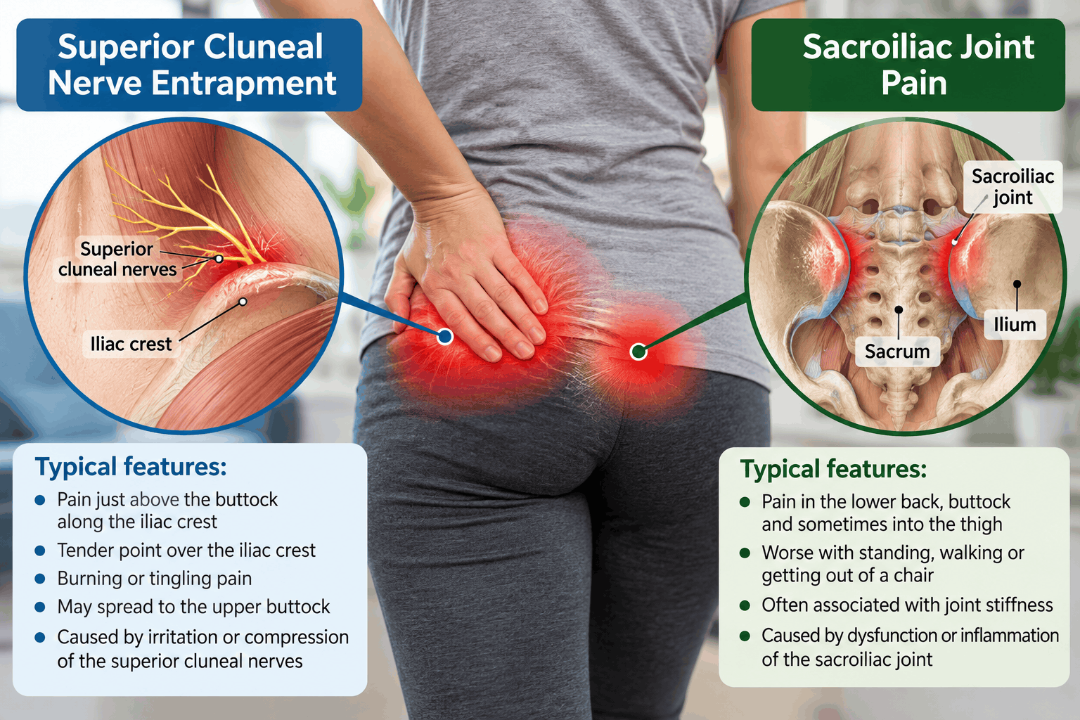
However, the two conditions affect neighboring anatomical structures, so their pain patterns frequently overlap.
Pain Location in Superior Cluneal Nerve Entrapment
Superior cluneal nerve entrapment commonly produces pain along the posterior iliac crest.
This is the upper rim of the pelvic bone that can be felt beneath the skin at the back of the waist.
A characteristic tender point is often found approximately 7 centimeters from the midline of the lower back, although the precise location varies with individual anatomy and the affected nerve branch.
Patients may describe pain that begins near this point and spreads downward into the upper buttock.
The discomfort can feel sharp, burning, aching, or occasionally electric.
In some cases, pain extends into the thigh, making the condition resemble lumbar radiculopathy or sciatica.
One particularly useful clue is that pressing directly over the suspected nerve entrapment point reproduces the patient’s familiar symptoms.
Research describing the diagnostic features of superior cluneal nerve entrapment emphasizes localized tenderness over the posterior iliac crest, reproduction of radiating symptoms during compression, and improvement following a targeted nerve block. (2)
Pain Location in Sacroiliac Joint Dysfunction
Sacroiliac joint pain is often concentrated slightly lower and closer to the sacrum.
Patients frequently point to an area immediately below and near the posterior superior iliac spine.
This location is sometimes identified during a clinical assessment using the Fortin finger test.
In this examination, the patient indicates the area where pain is most consistently experienced.
Pain concentrated near the characteristic sacroiliac joint region may support the diagnosis, although the location alone cannot confirm it.
Sacroiliac joint pain can also spread into the buttock, groin, or thigh.
Some patients experience discomfort extending farther down the leg, which can complicate the distinction from nerve-related conditions.
The Practical Difference in Pain Location
A patient who can identify a sharply tender point directly along the upper posterior iliac crest may have superior cluneal nerve entrapment.
A patient whose pain feels deeper and is concentrated immediately around the posterior sacroiliac region may have sacroiliac joint-related pain.
However, these are clinical clues rather than definitive rules.
Pain referral patterns vary, and patients can have more than one source of pain.
How Do the Symptoms of Superior Cluneal Nerve Entrapment Differ From Sacroiliac Joint Pain?
Although both conditions can cause persistent lower back and buttock pain, certain symptom characteristics may help guide the diagnosis.
1. Burning or Tingling Pain May Suggest Superior Cluneal Nerve Entrapment
Superior cluneal nerve entrapment can produce symptoms associated with irritation of a sensory nerve.
Patients may describe burning, tingling, shooting pain, or an uncomfortable sensation when the skin over the affected area is touched.
Some report that pressing a specific spot along the pelvic rim sends pain into the buttock.
These symptoms are particularly suggestive when they occur together with a characteristic tender point.
Sacroiliac joint pain is more commonly described as a deep, aching, or mechanical discomfort.
Nevertheless, pain quality alone is not sufficiently reliable to distinguish the conditions.
2. Pain During Position Changes May Suggest Sacroiliac Joint Pain
Patients with sacroiliac joint pain may experience discomfort when rising from a chair, climbing stairs, rolling over in bed, or standing with more weight on one leg.
These activities can place additional mechanical stress across the pelvis.
However, superior cluneal nerve entrapment can also worsen with movement.
In particular, lumbar bending, extension, prolonged standing, and walking may aggravate an entrapped superior cluneal nerve.
Therefore, pain during movement does not automatically indicate sacroiliac joint dysfunction.
3. A Small, Highly Sensitive Spot Favors Superior Cluneal Nerve Entrapment
One of the more distinctive findings in superior cluneal nerve entrapment is a localized trigger point over the posterior iliac crest.
The patient may be able to identify the painful spot with one finger.
When the clinician presses this point, the patient may experience a familiar shooting or radiating sensation.
This response is sometimes described as a Tinel-like sign.
In sacroiliac joint pain, tenderness may be present around the posterior joint region, but a characteristic nerve-related response at the iliac crest is less typical.
4. Pain Radiating Into the Leg Can Occur With Either Condition
Pain extending into the buttock or leg is often assumed to indicate a lumbar disc problem.
However, both superior cluneal nerve entrapment and sacroiliac joint disorders can produce referred leg pain.
A review of superior and middle cluneal nerve entrapment reported leg symptoms in a substantial proportion of affected patients. This explains why cluneal nerve disorders can resemble lumbar radiculopathy. (2)
The presence of radiating pain should therefore prompt a careful neurological and musculoskeletal examination rather than an immediate assumption that the problem originates from a spinal nerve root.
Progressive weakness, significant sensory loss, or abnormal reflexes require evaluation for other neurological causes.
How Can Doctors Tell the Difference During a Physical Examination?
A careful physical examination is often the most important starting point.
The clinician generally evaluates the location of pain, tenderness, lumbar spine movement, neurological findings, and responses to specific examination maneuvers.
Neither condition should be diagnosed solely from one positive physical examination finding.
Examining the Posterior Iliac Crest for Superior Cluneal Nerve Entrapment
When superior cluneal nerve entrapment is suspected, the clinician palpates along the posterior iliac crest.
The examination focuses on identifying a localized point of maximal tenderness where the nerve may cross the pelvic rim.
Pressure over this area may reproduce the patient’s usual lower back pain.
A particularly suggestive response occurs when pressure produces tingling, numbness, or radiating discomfort in the distribution of the superior cluneal nerve.
This is often called a positive Tinel-like sign.
The clinician may also examine sensation over the upper buttock and assess lumbar movements that reproduce symptoms.
The combination of a characteristic tender point and reproduction of familiar radiating pain increases suspicion for superior cluneal nerve entrapment.
However, local tenderness alone is not enough to establish the diagnosis.
The Fortin Finger Test for Sacroiliac Joint Pain
The Fortin finger test is used to assess whether the patient’s pain is concentrated in a location commonly associated with the sacroiliac joint.
The patient is asked to point to the area of greatest discomfort.
Pain localized near the posterior superior iliac spine, particularly just below and toward the sacrum, may support sacroiliac joint involvement.
This finding becomes more meaningful when considered alongside other examination results.
A positive Fortin finger test does not independently prove that the sacroiliac joint is the pain generator.
Sacroiliac Joint Provocation Tests
Clinicians commonly use several physical examination maneuvers to determine whether mechanical stress across the sacroiliac region reproduces the patient’s familiar pain.
These include:
- Thigh thrust test: Pressure is transmitted through the femur toward the pelvis to stress the sacroiliac region.
- Sacroiliac joint compression test: Pressure is applied across the pelvis while the patient lies on the side.
- Sacroiliac joint distraction test: Pressure is applied to the front of the pelvis to stress the sacroiliac structures.
- Gaenslen’s test: The hips are positioned to create opposing forces across the pelvis.
- Patrick’s test: The hip is placed into flexion, abduction, and external rotation to assess whether the maneuver reproduces familiar pain.
Historically, three or more positive tests within an established cluster have been used to increase suspicion for sacroiliac joint pain.
However, more recent evidence indicates that positive provocation tests are less reliable for confirming sacroiliac joint pain than negative clusters are for making the diagnosis less likely.
A 2025 multispecialty consensus guideline emphasized that no individual historical feature or physical examination maneuver can reliably establish sacroiliac joint pain.
This is why diagnostic injections may be considered when symptoms persist and the source remains uncertain.
Diagnostic Nerve Block vs Sacroiliac Joint Injection: Which Test Is More Useful?
When physical examination findings overlap, targeted diagnostic injections may help identify the source of pain.
The important distinction is that these procedures target different anatomical structures.
Superior Cluneal Nerve Block for Suspected Nerve Entrapment
A superior cluneal nerve block involves injecting a local anesthetic near the suspected nerve entrapment site.
The injection may be performed using anatomical landmarks or ultrasound guidance, depending on the clinical circumstances.
The goal is to temporarily interrupt pain signals from the superior cluneal nerve.
If the patient’s familiar pain decreases substantially during the expected period of anesthetic activity, this supports the possibility that the superior cluneal nerve is contributing to the symptoms.
For example, a patient with persistent pain along the posterior iliac crest may experience marked improvement after a targeted nerve block.
Such a response becomes particularly meaningful when the injection corresponds with the characteristic tender point identified during examination.
However, a nerve block is not infallible.
Local anesthetic can spread into neighboring tissues, and the degree of relief may depend on injection placement, technique, and the presence of other pain generators.
A negative response also does not completely exclude nerve entrapment.
Diagnostic Sacroiliac Joint Injection
A diagnostic sacroiliac joint injection is performed to determine whether pain originates from structures inside the joint.
Using imaging guidance, a clinician places a needle into the sacroiliac joint and injects a small amount of local anesthetic.
If the patient’s usual pain decreases significantly during the anesthetic’s expected duration, the result supports an intra-articular sacroiliac pain source.
However, pain can also originate from ligaments and other structures outside the joint cavity.
An injection into the joint may not adequately assess all of these structures.
Diagnostic approaches for extra-articular sacroiliac pain can therefore differ from those used for pain originating within the joint.
Current clinical guidelines emphasize that the interpretation of diagnostic injections depends on the suspected anatomical source, injection technique, degree of relief, and the treatment being considered.
What If a Sacroiliac Joint Injection Does Not Relieve the Pain?
An unsuccessful sacroiliac joint injection does not automatically establish superior cluneal nerve entrapment.
However, it should encourage reconsideration of the diagnosis.
If the patient continues to have a sharply localized tender point over the posterior iliac crest, examining the superior cluneal nerve becomes particularly relevant.
Other possible explanations include lumbar spine disorders, gluteal tendon problems, myofascial pain, and middle cluneal nerve entrapment.
The response to a properly performed injection should always be interpreted together with the patient’s symptoms and examination findings.
Can a Normal MRI Scan Rule Out Superior Cluneal Nerve Entrapment?
No. A normal magnetic resonance imaging scan does not rule out superior cluneal nerve entrapment.
The superior cluneal nerves are small, and their entrapment is generally not identified on routine lumbar spine imaging.
This means a patient can have persistent nerve-related pain even when magnetic resonance imaging shows no significant disc herniation, spinal stenosis, or other obvious structural abnormality.
Similarly, imaging may not reliably identify the source of mechanical sacroiliac joint pain.
Magnetic resonance imaging can be valuable when inflammatory sacroiliitis, infection, fracture, malignancy, or another structural disorder is suspected.
However, it is less useful for determining whether an otherwise unremarkable sacroiliac joint is responsible for mechanical pain.
A normal scan should therefore be interpreted as one part of the diagnostic evaluation rather than proof that no physical cause of pain exists.
Research on cluneal nerve entrapment emphasizes the importance of clinical examination and targeted nerve blocks because conventional imaging frequently cannot demonstrate the affected nerve.
Can Superior Cluneal Nerve Entrapment Be Mistaken for Sacroiliac Joint Dysfunction?
Yes. Superior cluneal nerve entrapment can be mistaken for sacroiliac joint dysfunction because the painful regions are close together.
Both conditions can cause unilateral lower back pain, buttock discomfort, and symptoms aggravated by everyday movements.
A patient may initially receive a diagnosis of sacroiliac joint dysfunction based on pain location and mechanical symptoms.
If treatment provides little improvement, the clinician may need to investigate other structures around the posterior pelvis.
The superior cluneal nerve is one possibility.
Another important consideration is middle cluneal nerve entrapment.
Middle Cluneal Nerve Entrapment Can Also Mimic Sacroiliac Joint Pain
The middle cluneal nerves provide sensation to portions of the medial buttock.
Their anatomical course brings them close to the posterior sacroiliac ligaments.
When these nerves become entrapped, pain may occur in a location particularly similar to sacroiliac joint pain.
Unlike superior cluneal nerve entrapment, which commonly produces a tender point along the upper posterior iliac crest, middle cluneal nerve entrapment may produce tenderness lower down, near the posterior sacroiliac region.
A study published in Acta Neurochirurgica specifically examined how middle cluneal nerve entrapment can mimic sacroiliac joint-related pain.
The authors concluded that middle cluneal nerve entrapment should be considered when a patient suspected of having sacroiliac joint pain does not obtain satisfactory relief from a sacroiliac joint block.
This distinction matters because patients with persistent pain near the sacroiliac joint may have irritation involving the superior cluneal nerve, middle cluneal nerve, sacroiliac structures, or a combination of these.
Can You Have Superior Cluneal Nerve Entrapment and Sacroiliac Joint Pain at the Same Time?
Yes. The two conditions are not mutually exclusive.
The lower back and pelvis contain multiple potential sources of pain, including joints, ligaments, muscles, tendons, and sensory nerves.
A patient may have mechanical sacroiliac joint pain together with irritation of a nearby cluneal nerve.
This possibility becomes relevant when treatment directed at one structure provides only partial relief.
For example, a patient may experience improvement in deep buttock pain following a sacroiliac joint injection but continue to have a sharply tender area along the posterior iliac crest.
That remaining pain may require a separate evaluation.
Likewise, a superior cluneal nerve block may relieve localized burning pain while deeper mechanical discomfort persists.
Partial relief does not necessarily mean the original diagnosis was incorrect. It may indicate that more than one structure is contributing to the symptoms.
A thorough assessment should therefore consider the possibility of overlapping pain generators rather than assuming every symptom must originate from a single condition.
Treatment for Superior Cluneal Nerve Entrapment vs Sacroiliac Joint Pain
Treatment depends on the identified source of pain, symptom severity, duration, and response to previous care.
Although some conservative approaches overlap, the procedures used for persistent symptoms can differ substantially.
Treatment Options for Superior Cluneal Nerve Entrapment
Initial treatment may include activity modification, physical therapy, and medications selected according to the patient’s symptoms and medical history.
Physical therapy may focus on improving movement patterns, reducing mechanical irritation, and addressing contributing muscle dysfunction.
When symptoms persist, a superior cluneal nerve block may be considered.
This injection can serve both diagnostic and therapeutic purposes.
Some patients experience meaningful relief after a single injection, while others require additional treatment.
For persistent, well-localized nerve-related pain, specialists may consider radiofrequency treatment or other targeted interventions.
In carefully selected patients with refractory symptoms and convincing evidence of nerve entrapment, surgical decompression may be an option.
The evidence supporting some advanced treatments remains limited, and outcomes vary.
A comprehensive review of cluneal neuralgia describes nerve blocks, radiofrequency-based interventions, peripheral nerve stimulation, and surgical approaches as potential options for selected patients. (1)
Treatment Options for Sacroiliac Joint Pain
Conservative treatment for sacroiliac joint pain commonly includes physical therapy, activity modification, and appropriate pain-relieving medications.
Rehabilitation may emphasize pelvic stability, hip strength, trunk muscle coordination, and movement strategies that reduce symptom aggravation.
If conservative treatment does not provide adequate relief, an image-guided sacroiliac joint injection may be considered.
For selected patients with pain arising from the posterior sacroiliac joint complex, radiofrequency ablation targeting the relevant sacral lateral branches may provide longer-lasting relief.
In carefully selected patients with persistent, confirmed sacroiliac joint pain despite appropriate nonsurgical treatment, sacroiliac joint fusion may be considered.
However, surgery is not appropriate for most patients with pain near the sacroiliac joint.
The diagnosis must be carefully established before invasive treatment is pursued.
The American Society of Pain and Neuroscience provides recommendations addressing conservative care, diagnostic injections, radiofrequency ablation, and surgical treatment for appropriately selected patients with sacroiliac disorders. (3)
Why the Correct Diagnosis Matters Before Radiofrequency Ablation
Radiofrequency ablation is sometimes recommended for persistent lower back or sacroiliac region pain.
However, the success of the procedure depends heavily on identifying the appropriate pain-generating structure.
Radiofrequency treatment for superior cluneal nerve-related pain targets a different anatomical pathway from radiofrequency ablation used for posterior sacroiliac joint complex pain.
Treating the wrong structure may result in little or no meaningful improvement.
For example, a patient whose symptoms primarily originate from superior cluneal nerve entrapment may not benefit from a procedure directed at sacral lateral branches.
Similarly, treating the superior cluneal nerve may not adequately relieve pain originating from the sacroiliac joint.
This is why clinicians often rely on a combination of clinical findings and appropriately selected diagnostic blocks before recommending certain interventional procedures.
Patients should understand which structure is being targeted, why it is suspected to be responsible for their pain, and what evidence supports the proposed treatment.
What Should You Do If Sacroiliac Joint Treatment Has Not Worked?
Persistent pain after treatment for suspected sacroiliac joint dysfunction deserves reassessment.
It does not necessarily mean the pain is untreatable or that the patient needs increasingly invasive procedures.
Instead, the original diagnosis may need to be reconsidered.
A clinician may review whether the pain is truly centered over the sacroiliac joint or whether the most tender area lies higher along the posterior iliac crest.
The examination may include palpation of the superior and middle cluneal nerve pathways, assessment of lumbar spine movement, neurological testing, and evaluation of surrounding muscles and tendons.
The clinician may also review previous injection reports to determine whether the intended anatomical structure was accurately targeted and whether the patient’s response was consistent with the expected anesthetic effect.
When examination findings suggest cluneal nerve entrapment, a targeted diagnostic nerve block may be appropriate.
Importantly, persistent lower back pain can also originate from lumbar facet joints, intervertebral discs, hip disorders, or other structures.
A comprehensive evaluation should not focus exclusively on the sacroiliac joint and cluneal nerves.
When Should Lower Back or Buttock Pain Be Evaluated Urgently?
Most cases of superior cluneal nerve entrapment and mechanical sacroiliac joint pain are not medical emergencies.
However, certain symptoms require prompt medical attention because they may indicate a more serious condition.
Seek emergency evaluation for new urinary retention, loss of bladder or bowel control, numbness around the groin or saddle region, or rapidly progressive leg weakness.
These symptoms can indicate significant neurological compression.
Prompt medical assessment is also important when back pain occurs with fever, unexplained weight loss, a history of cancer, significant trauma, or other concerning systemic symptoms.
Severe pain following a fall, particularly in someone with osteoporosis, may require evaluation for a fracture.
These warning signs should not be attributed to cluneal nerve entrapment or sacroiliac joint dysfunction without appropriate investigation.
Frequently Asked Questions About Superior Cluneal Nerve Entrapment and Sacroiliac Joint Pain
Can superior cluneal nerve entrapment feel exactly like sacroiliac joint pain?
Yes. Both conditions can produce pain in the lower back and buttock, particularly on one side. Superior cluneal nerve entrapment may be more likely when there is a sharply localized tender point along the posterior iliac crest, especially if pressure causes tingling or radiating pain. However, symptoms alone cannot reliably distinguish the conditions.
Where do you press to check for superior cluneal nerve entrapment?
Clinicians commonly examine the posterior iliac crest, particularly around 7 centimeters from the midline, while accounting for anatomical variation. Reproduction of familiar radiating pain or tingling may support the diagnosis. Patients should not attempt forceful self-examination because tenderness in this region can have several causes.
Is superior cluneal nerve entrapment the same as sacroiliac joint dysfunction?
No. Superior cluneal nerve entrapment involves irritation or compression of a sensory nerve crossing the posterior iliac crest. Sacroiliac joint dysfunction refers to pain associated with the joint between the sacrum and pelvis or its supporting structures. The conditions can produce similar symptoms but may require different treatments.
Can superior cluneal nerve entrapment cause pain when sitting?
Yes. Some patients experience worsening discomfort during prolonged sitting, particularly when posture or pressure increases irritation around the affected nerve. However, sitting-related pain is not specific to cluneal nerve entrapment and may also occur with sacroiliac joint disorders, lumbar spine conditions, and other causes of buttock pain.
Can sacroiliac joint pain cause burning sensations?
Yes. Although sacroiliac joint pain is commonly described as deep and aching, patients may report burning or radiating discomfort. Pain quality alone cannot reliably identify the source. Burning accompanied by localized tingling or a Tinel-like response over the posterior iliac crest may raise suspicion for cluneal nerve involvement.
Will a superior cluneal nerve block confirm the diagnosis?
A substantial reduction in familiar pain following a correctly targeted superior cluneal nerve block supports the diagnosis, particularly when characteristic examination findings are present. However, diagnostic injections are not perfectly specific, and results must be interpreted in the context of the overall clinical assessment.
Why does my sacroiliac joint hurt if my magnetic resonance imaging scan is normal?
Mechanical pain involving the sacroiliac joint or its supporting structures may occur without obvious abnormalities on routine imaging. Superior cluneal nerve entrapment may also cause pain despite a normal scan. Clinical examination and appropriately selected diagnostic procedures may help determine the source.
Can physical therapy help both conditions?
Physical therapy may help patients with either condition, particularly when muscle weakness, movement limitations, or altered mechanics contribute to symptoms. However, the rehabilitation approach may differ, and persistent symptoms should prompt reassessment rather than indefinite continuation of an ineffective treatment program.
Which specialist diagnoses superior cluneal nerve entrapment?
Pain management physicians, physiatrists, neurologists, and spine specialists may evaluate suspected superior cluneal nerve entrapment. Clinicians experienced in peripheral nerve disorders and image-guided diagnostic injections may be particularly helpful when the diagnosis remains uncertain.
The Bottom Line: Is the Pain Coming From the Nerve or the Sacroiliac Joint?
Superior cluneal nerve entrapment and sacroiliac joint pain can be difficult to distinguish because both affect the lower back and posterior pelvis.
The most useful clue for superior cluneal nerve entrapment is often a distinct tender point along the posterior iliac crest that reproduces familiar pain, tingling, or radiating discomfort when pressed.
Sacroiliac joint pain is more commonly associated with deeper discomfort near the posterior sacroiliac region and pain reproduced by a combination of mechanical provocation tests.
Neither finding is definitive on its own.
A targeted superior cluneal nerve block may help determine whether the nerve contributes to symptoms, while an appropriately performed sacroiliac joint injection may help identify pain originating from within the joint.
For patients who have undergone treatment for presumed sacroiliac joint dysfunction without meaningful improvement, superior cluneal nerve entrapment deserves consideration.
The goal is not simply to identify where the pain is felt, but to determine which anatomical structure is actually generating it.
That distinction can make a substantial difference in choosing an appropriate treatment and avoiding unnecessary procedures.
- Anderson D, et al. A Comprehensive Review of Cluneal Neuralgia as a Cause of Lower Back Pain. Orthopedic Reviews. 2022. https://pmc.ncbi.nlm.nih.gov/articles/PMC9235435/
- Isu T, Kim K, Morimoto D, Iwamoto N. Superior and Middle Cluneal Nerve Entrapment as a Cause of Low Back Pain. Neurospine. 2018;15(1):25–32. https://pmc.ncbi.nlm.nih.gov/articles/PMC5944640/
- American Society of Pain and Neuroscience Best Practice Guideline for the Treatment of Sacroiliac Disorders. Journal of Pain Research. 2024. https://pmc.ncbi.nlm.nih.gov/articles/PMC11075694/
- Consensus Practice Guidelines on Sacroiliac Joint Complex Pain From a Multispecialty, International Working Group. Regional Anesthesia & Pain Medicine. 2025. https://pmc.ncbi.nlm.nih.gov/articles/PMC12681192/
- Superior Cluneal Nerve Entrapment Syndrome: A Common but Often Overlooked Cause of Low Back Pain. 2022. https://pmc.ncbi.nlm.nih.gov/articles/PMC9062575/
- Matsumoto J, et al. Middle Cluneal Nerve Entrapment Mimics Sacroiliac Joint Pain. Acta Neurochirurgica. 2019;161(4):657–661. https://pubmed.ncbi.nlm.nih.gov/30830272/
- Prospective Study of Superior Cluneal Nerve Disorder as a Potential Cause of Low Back Pain and Leg Symptoms. Journal of Orthopaedic Surgery and Research. 2014. https://pmc.ncbi.nlm.nih.gov/articles/PMC4299373/
- Characteristics of Low Back Pain Due to Superior Cluneal Nerve Entrapment Neuropathy. 2019. https://pmc.ncbi.nlm.nih.gov/articles/PMC6773996/
Medical Disclaimer: This article is intended for educational purposes and should not replace professional medical evaluation, diagnosis, or treatment. Persistent or worsening lower back pain should be assessed by a qualified healthcare professional.